Induced pluripotent stem cell-derived (iPSC) organoids offer unprecedented opportunities to study human development, disease, and drug responses in a controlled in vitro environment.
These 3D models mimic the functionality of human organs and new methodologies are being rapidly developed to study their complex physiological structure and processes.
Gain deep insights into 3D human in vitro organoid models with label-free, live-cell assays from Axion BioSystems.
Advantages iPSC organoid models
Organoids are a powerful tool for biological research.
Advantages of iPSC organoids:
- Functional similarity
iPSC organoids recapitulate complex physiological processes seen in vivo. - Self organizing
iPSC organoids self-organize into 3D structures similar to tissues in vivo. - Cellular diversity
iPSC organoids are often comprised of multiple cell types found in their target organ (e.g. neurons & glial cells).
With increased physiological relevance organoids are used for applications in drug screening, disease modeling, and personalized medicine.

Assays for iPSC organoid characterization and function
-
Standardizing neural organoid production and analysis>
-
Neural organoids model development and maturation of brain networks>
-
Cardiac organoids to model ischemia-reperfusion injury>
-
Label-free tracking of hepatic organoids in vitro>
-
Publication Highlight: Live-cell imaging with 3D models>
-
Additional Resources: Neural Organoids>
-
Publications: Cardiac Organoids>
Quality neural organoid research requires the right tools from start to finish.
Watch this 8 minute video to discover how Axion's imaging and MEA platforms allow you to capture the complex biology of organoids non-invasively and in real time.
Purpose: To model human brain development with cortical organoids. Altered brain development in utero can have lifelong consequences but a lack of appropriate in vitro models limits our ability to investigate these processes.
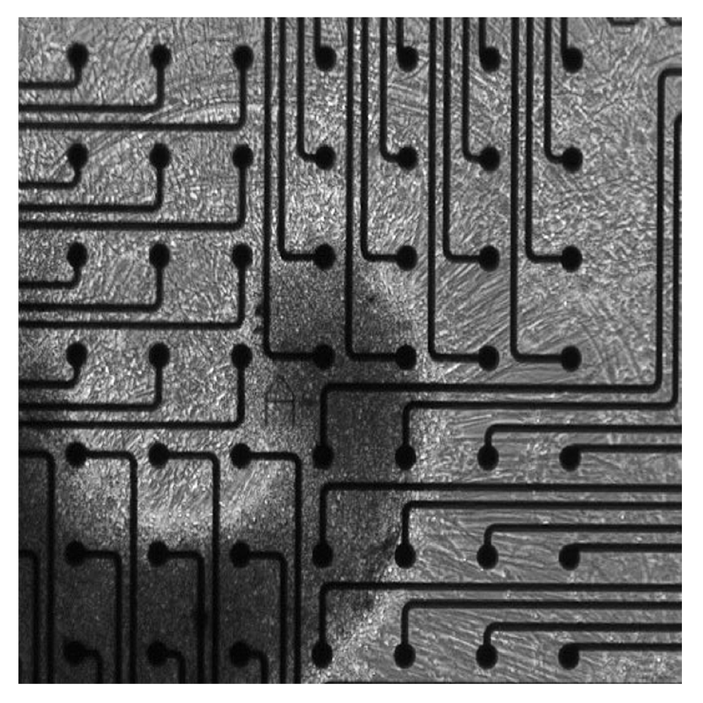
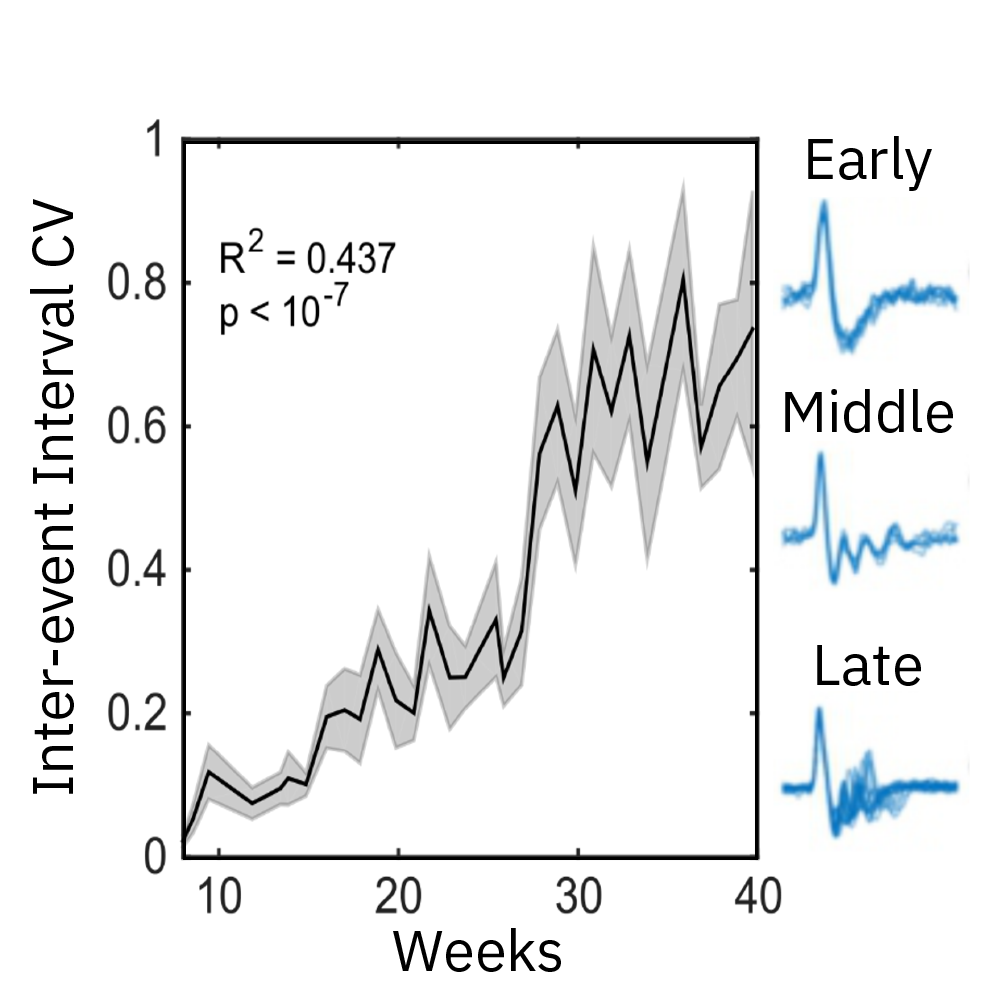
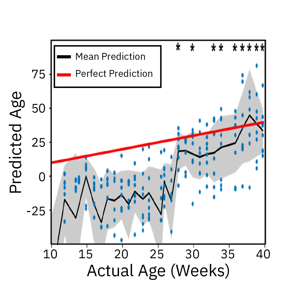
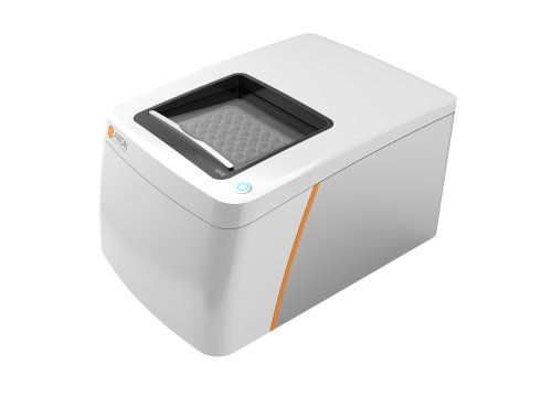
Extracellular recordings of spontaneous electrical activity of neural organoids were measured using the Maestro MEA platform over the course of 10 months and compared to neonatal EEGs.
Result: Cortical organoid activity demonstrated a continuously evolving neural network growing in complexity from early, middle and late time points. Organoid maturation and development were found to mimic the development of the preterm neonatal brain. Watch this webinar to learn more.
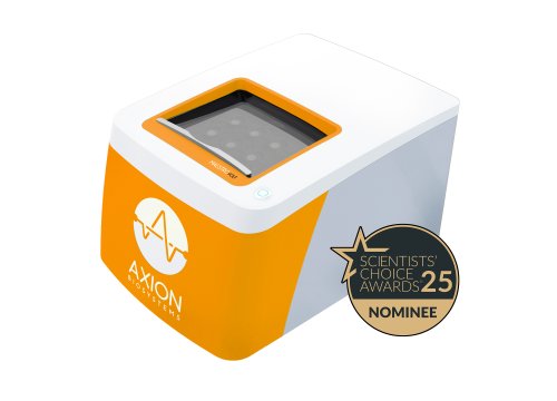
Ischemia-reperfusion injury, a primary driver of cardiac disease, is known to affect more than cardiomyocytes alone. Multicellular cardiac organoids provide a more clinically-relevant in vitro model to investigate the complex interactions between the cells of the heart and their implications in disease.
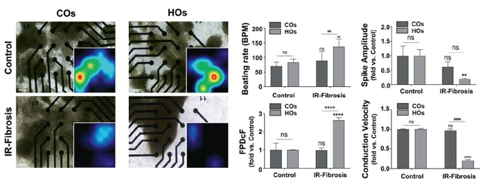
To demonstrate the importance of multicellular interactions in ischemia-reperfusion injury in vitro, and the electrophysiological defects associated with it, cardiac organoids (COs) comprised of cardiomyocytes alone and heart organoids (HOs) comprised of cardiomyocytes, fibroblasts, and endothelial cells were created. Maestro MEA was used to measure activity following ischemia-reperfusion injury.
Results: 3D multicellular heart organoids were an effective model of ischemia-reperfusion injury. Both cardiac organoids and heart organoids led to electrophysiological alterations, but multicellular heart organoids demonstrated a far more robust response. A two-fold increase in field potential duration (Fridericia-corrected) and a significant slowing of conduction velocity was present in the heart organoids, indicative of an elevated risk of arrhythmogenesis. Data from Song et al, 2024, provided by NEXEL.
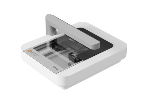
The liver plays a vital role in drug metabolism. Induced pluripotent stem cell-derived hepatic organoids mimic liver structure and function and are a valuable tool for understanding drug-induced liver injury (DILI) and disease. The Omni platform can be used to monitor the growth and development of iPSC-derived hepatic organoids.
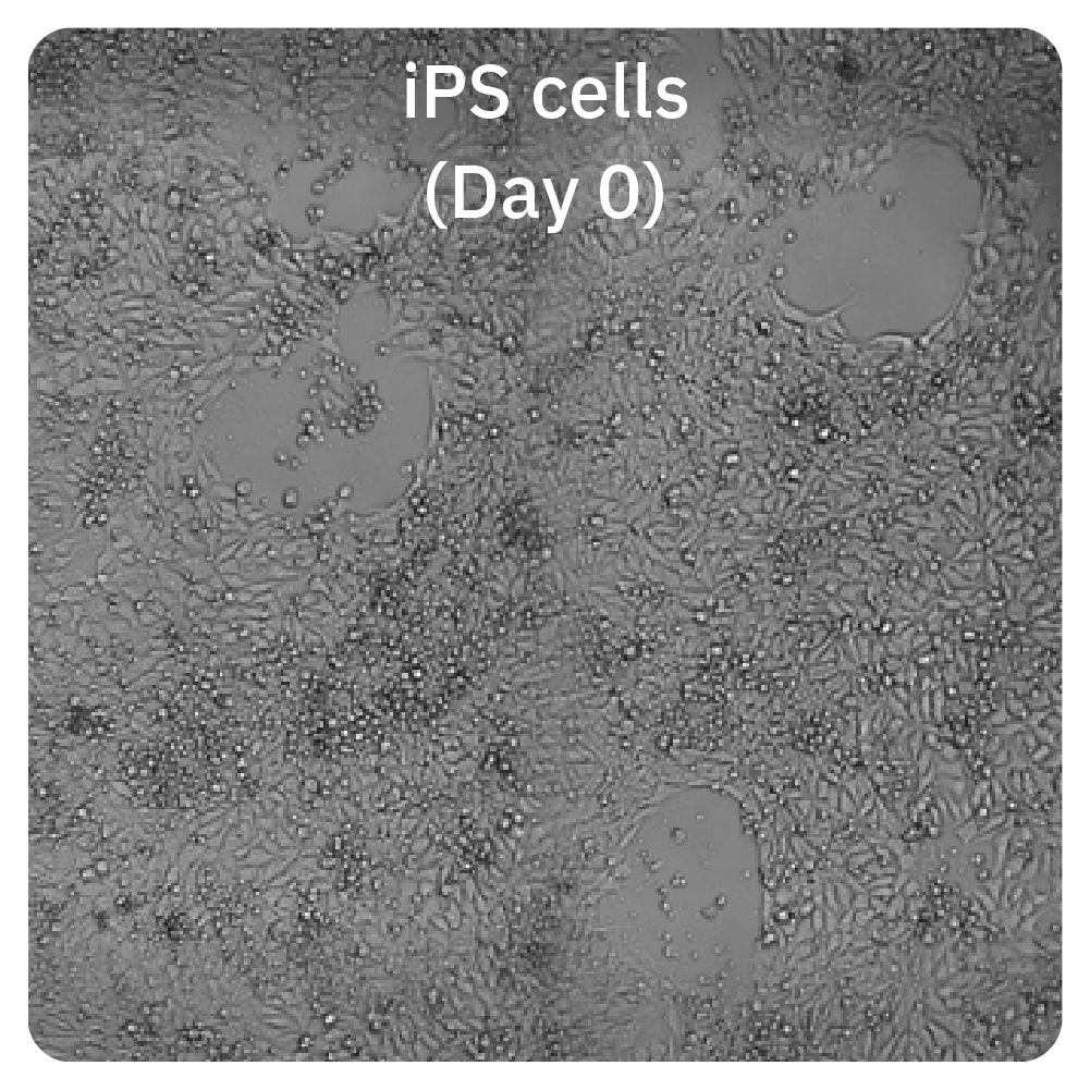
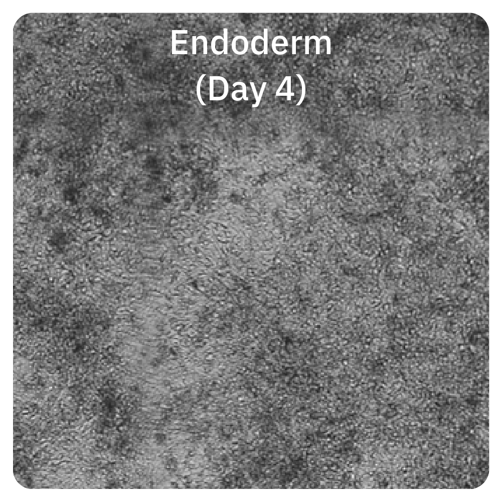
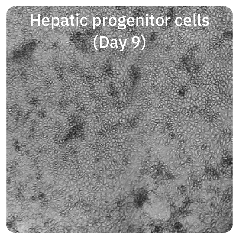
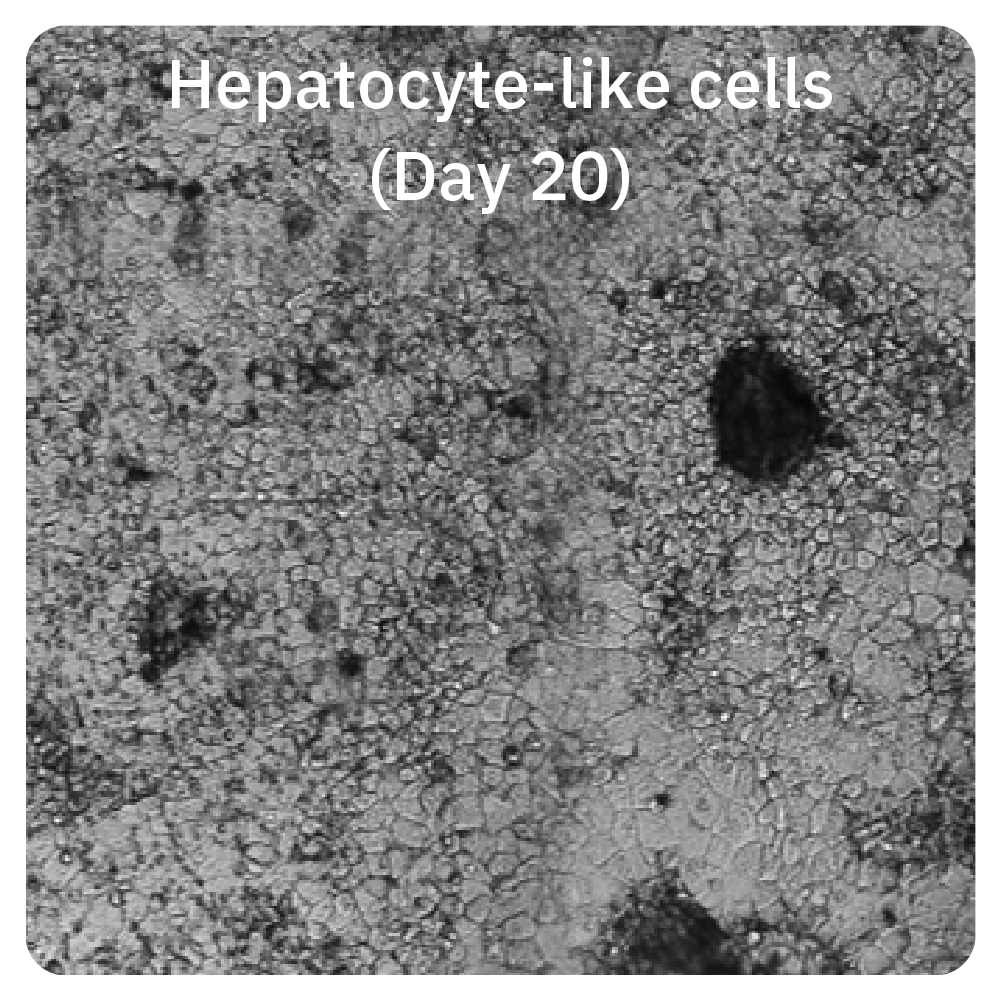
iPS cells were imaged on the Omni during differentiation and the expected morphological changes were observed during the development of hepatocytes.
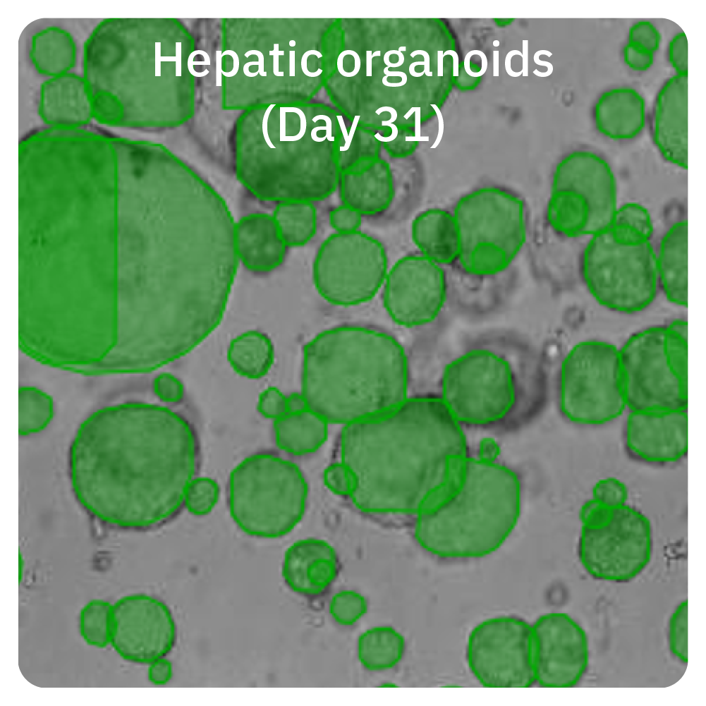
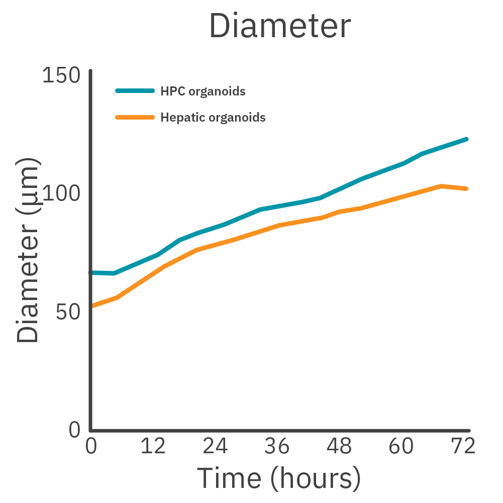
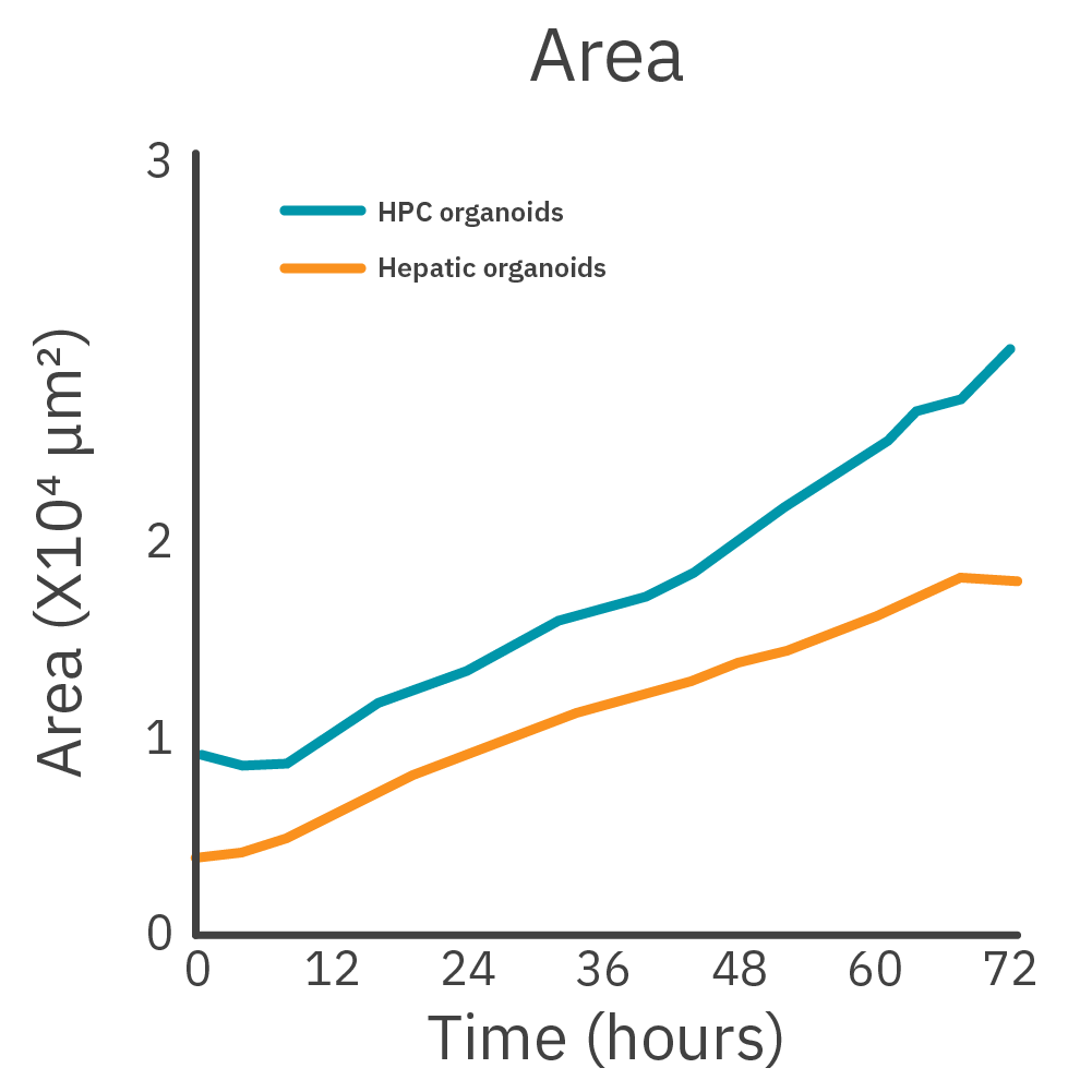
During differentiation, the hepatic progenitor cells and hepatocyte-like cells were transferred to organoid culture. The Omni was used to measure organoid growth and development.
Live-cell imaging on the Omni platform can be used to monitor iPSC-derived hepatic organoids.
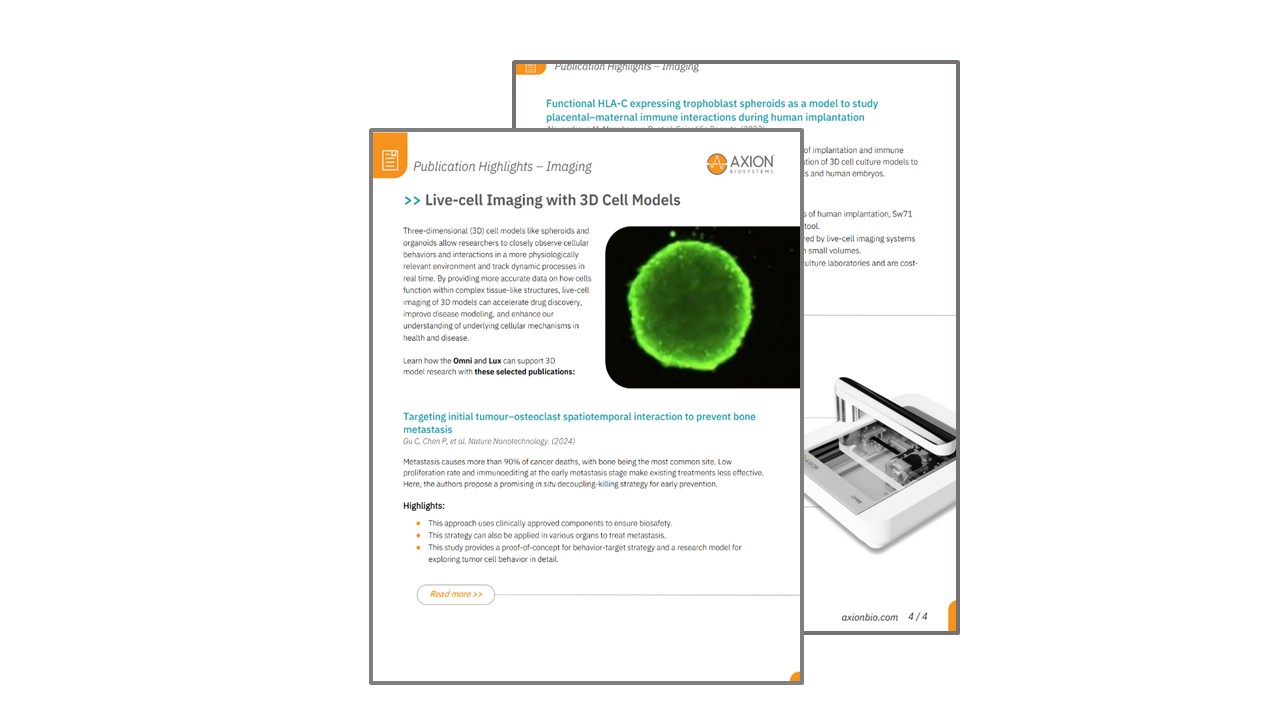
By providing more accurate data on how cells function within complex tissue-like structures, live-cell imaging of 3D models can accelerate drug discovery, improve disease modeling, and enhance our understanding of underlying cellular mechanisms in health and disease. Learn how the Omni and Lux live-cell imaging platforms can support 3D model research with these selected publications.
See how iPSC-derived neural organoids and our next-generation instrument platforms are transforming neuroscience research
Featured on this page:
>> Coffee Break Webinars
>> PBS Documentary: The Cannabis Question
>> Publications
>> Application Notes and Culture Protocols
>> In the News
Three-dimensional cardiac organoids offer more physiologically relevant in vitro cell models of the human heart, enabling scientists to better mimic the complex structure and cellular environment found in vivo.
Discover how researchers are using 3D cardiac organoids on Axion BioSystems’ hands-free Maestro MEA platform to improve disease modeling, accelerate drug screening, reduce reliance on animal models, and facilitate the development of novel therapeutic approaches for heart disease.