
Clonogenic Assay Module
Automatically detect, count, and measure colonies with whole-well live-cell imaging. The Clonogenic Assay Module for Omni helps researchers quantify colony formation over time, reducing manual counting while providing longitudinal measurements of colony number, size, and morphology.
Key Features
Automate clonogenic assay imaging and analysis

Count colonies across the whole well
Whole-well imaging captures colonies across the entire culture area, helping reduce sampling bias in low-density clonogenic assays.
Monitor colony formation over time
Automatically image cultures at defined intervals to follow colony emergence, growth, and treatment response over days or weeks.

Reduce manual variability
Replace time-consuming manual colony counting with automated detection and consistent image analysis.
Quantify colony growth and morphology
Measure colony count, size, and circularity to compare experimental conditions and treatment effects.
Go beyond endpoint colony counting
Traditional clonogenic assays are often evaluated at a single endpoint after colonies have formed. But colony formation is highly time dependent: colonies emerge, expand, and can eventually merge.
With the Clonogenic Assay Module, Omni repeatedly images the same cultures throughout the experiment, allowing researchers to see when colonies form, how quickly they grow, and how experimental treatments change colony development over time.
Whole-well imaging also helps ensure that colonies distributed across the culture vessel are included in the analysis rather than relying on selected fields of view.
Overview
What can the Clonogenic Assay Module measure?

Colony count
Automatically detect and quantify the number of colonies within the imaged well.
Colony size
Measure average colony area to quantify changes in colony growth between conditions.
Circularity
Characterize colony shape and morphological changes during colony formation.
Colony formation over time
Follow changes in colony number and morphology across sequential imaging timepoints.
Whole-well imaging for more representative colony counts
Clonogenic assays are typically performed at low cell densities, where colonies may be distributed throughout the well. Imaging only selected fields of view can miss colonies and introduce sampling variability.
Omni captures the whole well so colonies throughout the culture area can be detected and analyzed.
- ✓ Analyze colonies across the entire well
- ✓ Follow colony formation longitudinally
- ✓ Reduce dependence on manual colony counting
- ✓ Compare treatment conditions quantitatively
Follow the kinetics of clonogenic survival
A single endpoint can tell you how many colonies remain at the end of an experiment. Longitudinal imaging provides additional information about how the response develops.
>> Early colony formation
Detect emerging colonies as surviving cells begin to proliferate.
>> Colony growth
Measure changes in colony number and size as cultures develop.
>> Treatment response
Compare the effects of cytotoxic agents, immunotherapies, radiation, or other experimental treatments on colony formation.
>> Optimal endpoint selection
Review colony growth over time to identify an appropriate analysis window before colonies become overly confluent or begin to merge.
From whole-well images to quantitative results
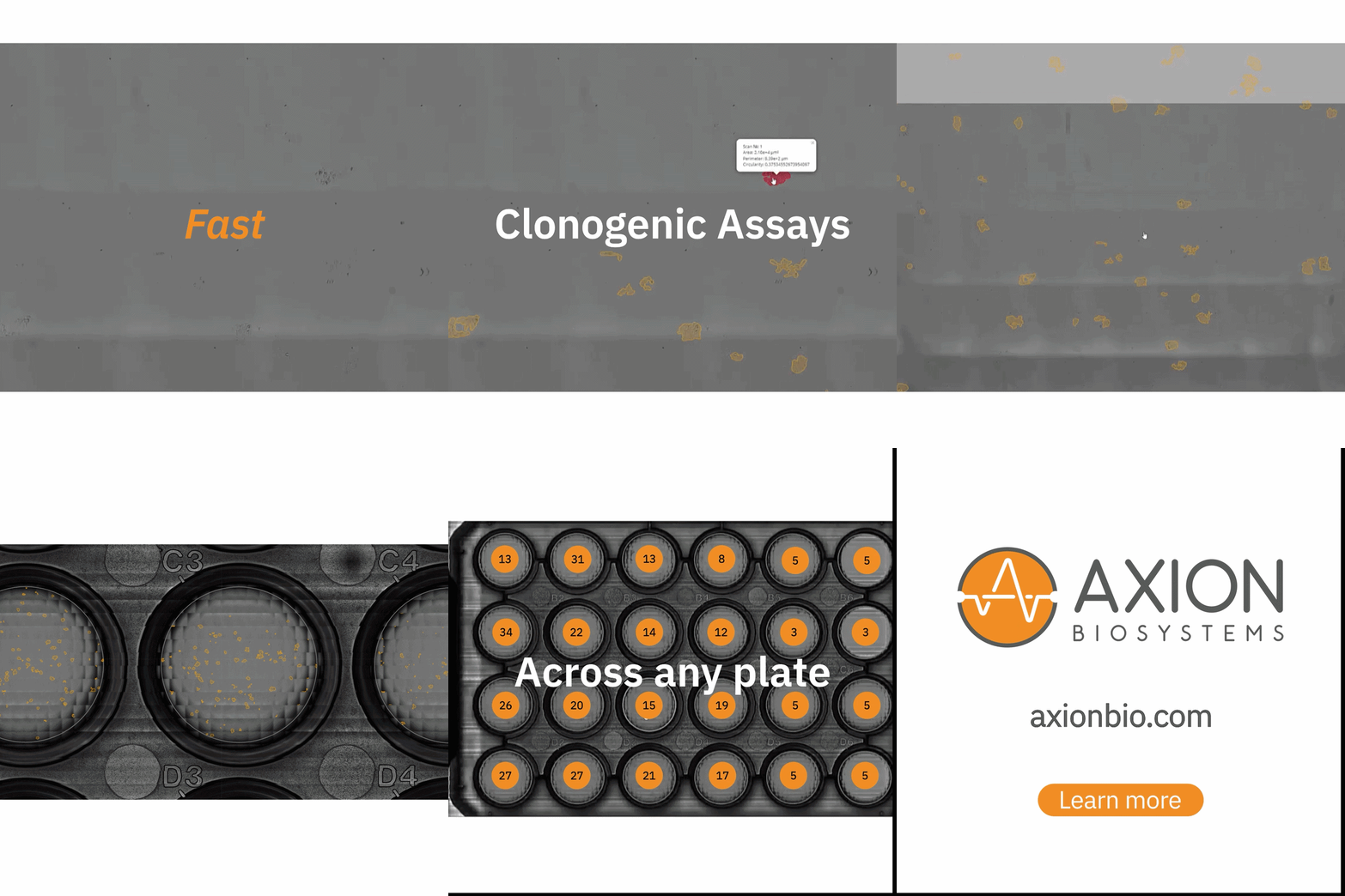
A single endpoint can tell you how many colonies remain at the end of an experiment. Longitudinal imaging provides additional information about how the response develops.
- 1. Automated imaging
Schedule repeated imaging without removing cultures from the incubator. - 2. Colony detection
Automatically identify colonies across the whole well. - 3. Quantitative analysis
Measure colony count, size, and circularity. - 4. Review images and results
Visualize colony masks, compare conditions, and review measurements through the Axion Portal.
Clonogenic assay applications
Use automated colony analysis to investigate cell survival and proliferative capacity across a range of experimental workflows.
Cancer drug response
Measure dose-dependent effects of anticancer compounds on colony formation and growth.
Cytotoxicity studies
Evaluate whether exposed cells retain the ability to survive and proliferate after treatment.
Radiation studies
Monitor colony formation following radiation exposure to evaluate clonogenic survival.
Cell and gene therapy research
Compare colony-forming capacity across experimental conditions, treatments, or engineered cell populations.
Stem cell workflows
Monitor colony formation during stem cell culture and clonal expansion workflows.
Frequently asked questions about the Clonogenic Assay Module
The Clonogenic Assay Module automatically detects and measures colonies in cultures imaged with Omni. Researchers can quantify colony count, size, and circularity while following colony formation over time.
Yes. The module automatically identifies colonies in the imaged culture area and calculates the number of colonies detected within each well.
Clonogenic assays are commonly performed at low cell densities, so colonies can be distributed throughout the well. Whole-well imaging captures the complete culture area rather than relying on selected fields of view, helping reduce sampling bias.
Yes. Omni can repeatedly image cultures at defined intervals, allowing researchers to follow colony emergence, growth, and changes in morphology throughout the experiment.
The module quantifies colony count, colony size or area, and colony circularity. These measurements can be compared across treatments, concentrations, and experimental conditions.
The module automates colony detection and measurement, reducing the need to manually identify and count colonies. Images and colony-detection overlays can also be reviewed to verify analysis results.
Yes. Clonogenic assays are commonly used to assess whether cells retain the ability to survive and proliferate after treatment. Automated longitudinal imaging can be used to compare colony formation across drug concentrations or treatment conditions.
Colony number and morphology change throughout an experiment as colonies form, expand, and eventually merge. Longitudinal imaging provides information about when these changes occur and can help researchers identify an appropriate analysis timepoint.
Colony formation can be monitored using brightfield imaging, allowing routine colony detection and morphology measurements without requiring fluorescent labels.
For Research Use Only. Not for use in diagnostic procedures.


