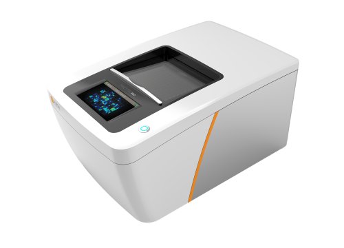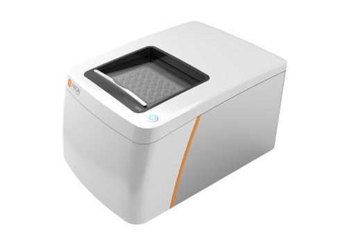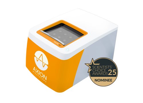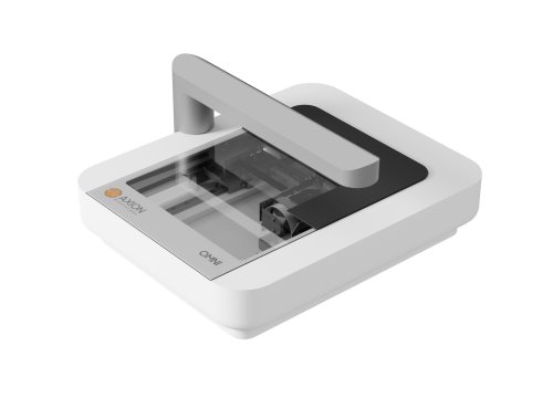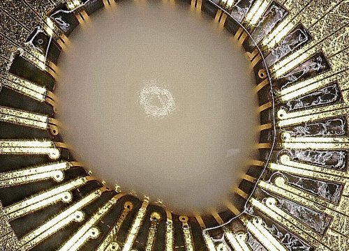
Measure neural activity, network development, and functional maturation in 3D neural models with noninvasive, real-time electrophysiology.
Neural organoids model key aspects of human brain development and function but can be challenging to measure functionally. The Maestro MEA platform enables longitudinal recording of neural activity from organoids, while Axion’s organoid-specific MEA plates are designed to improve placement, electrode contact, and signal detection in 3D models.
Functional Analysis of Neural Organoids
-
Standardizing organoid production and analysis>
-
Measure neural spiking and local field potentials simultaneously >
-
Following cortical organoid development over months>
-
Studying neuroimmune interactions in cortical organoids>
-
Neanderthal mini-brains>
-
Recording functional activity from cerebral organoids>
Quality neural organoid research requires the right tools from start to finish.
Watch this 8 minute video to discover how Axion's imaging and MEA platforms allow you to capture the complex biology of organoids non-invasively and in real time.
Organoids contain complex neuronal populations that can generate electrical activity at different spatial and temporal scales.
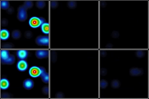
In the example shown on this page, neural organoids were recorded using the Maestro MEA platform. Neural spike activity was detected from the organoid.
Local field potentials, however, were detected in regions where surface spiking was not observed.
The ability to record both signals simultaneously provides complementary information about activity within the model.

What is the difference between spikes and local field potential?
Neural spikes represent rapid extracellular signals associated with neuronal action potentials.
Local field potentials are lower-frequency extracellular signals that reflect coordinated population-level electrical activity.
In a 3D organoid, combining these measurements can provide a broader picture of functional network activity than spike measurements alone.
One of the major advantages of noninvasive MEA recording is the ability to follow the same experimental model throughout development.
In the cortical organoid study highlighted on this page, spontaneous extracellular electrical activity was recorded over the course of 10 months and compared with neonatal EEG recordings.
Organoid network activity continued to evolve over time, with increasingly complex patterns appearing from early through later stages of development.
The analysis found that aspects of cortical organoid maturation resembled developmental features observed in the preterm neonatal brain.
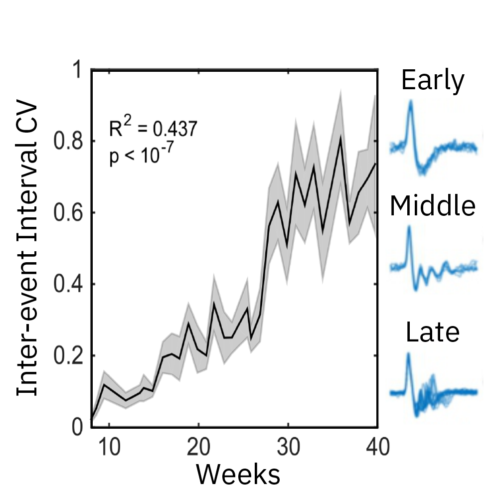
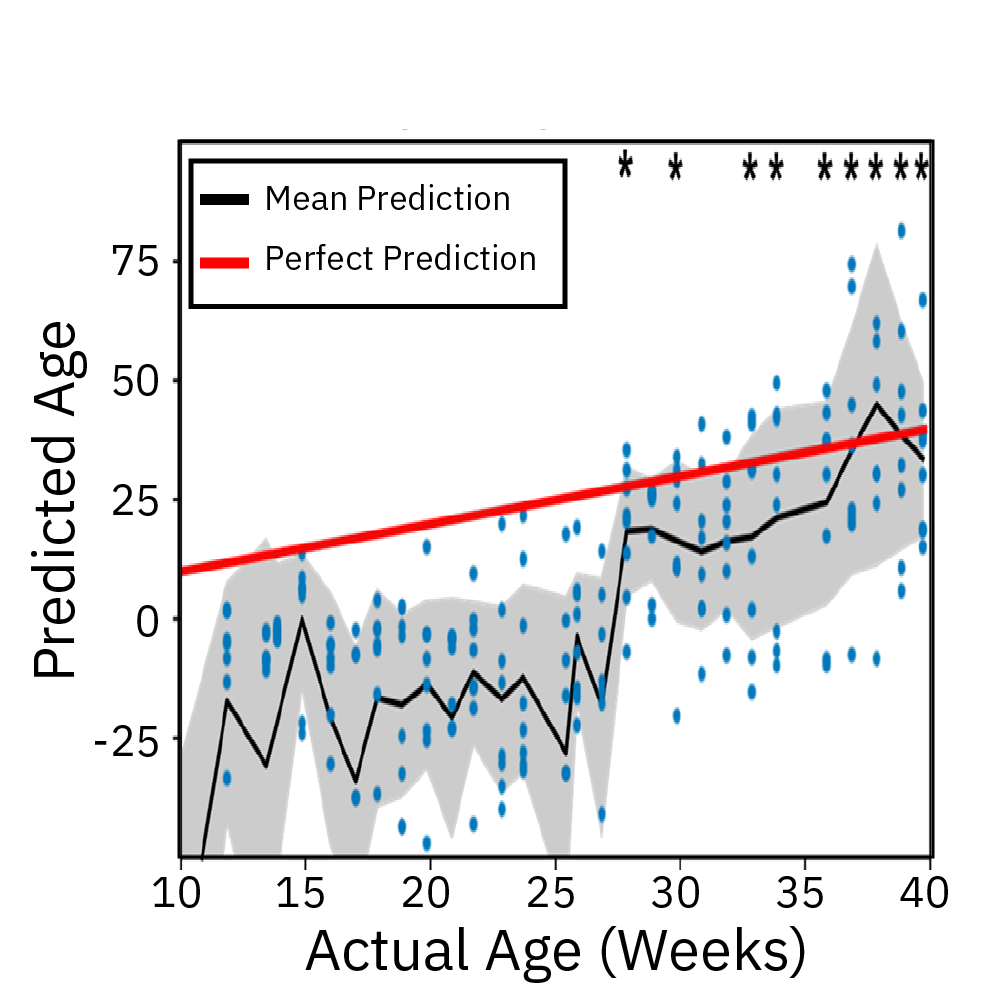
Spontaneous electrical activity from cortical organoids was recorded longitudinally using the Maestro MEA platform over 10 months. Network activity evolved in complexity across early, middle, and later developmental stages and demonstrated electrophysiological features associated with maturation.
Why longitudinal measurement matters
Organoid maturation can occur over much longer timescales than conventional neural cultures.
Repeated MEA recordings enable researchers to investigate:
- when spontaneous activity emerges;
- when coordinated network activity develops;
- how network complexity changes over time;
- whether functional maturation stabilizes;
- how different organoid batches compare;
- how disease models or treatments alter developmental trajectories.
The same model can be followed repeatedly without sacrificing the organoid at each measurement timepoint.
Neural organoids can incorporate multiple cell types to model more complex interactions within the nervous system.
Microglia, for example, play important roles in neural development and are implicated in neurological and neuropsychiatric disease.
In Popova et al, Cell Stem Cell 2021, researchers compared human microglia across culture models and used Maestro Pro MEA recordings to evaluate their functional effects after transplantation into cortical organoids.
The presence of microglia accelerated the emergence of synchronous, oscillatory network activity.
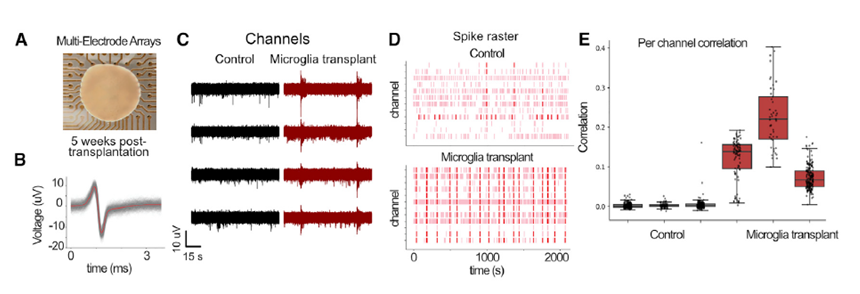
Cortical organoids on multiwell Maestro MEA system
The genomes of Neanderthals and modern humans are very similar. By investigating the differences in genetic make-up, we can gain insights into what separated modern humans from our extinct relatives. In this webinar, Cleber Trujillo, PhD (StemoniX) discusses how he introduced an archaic variant gene, NOVA1, into human pluripotent stem cell-derived brain organoids and evaluated the impact on neural activity as recorded on the Maestro MEA system.
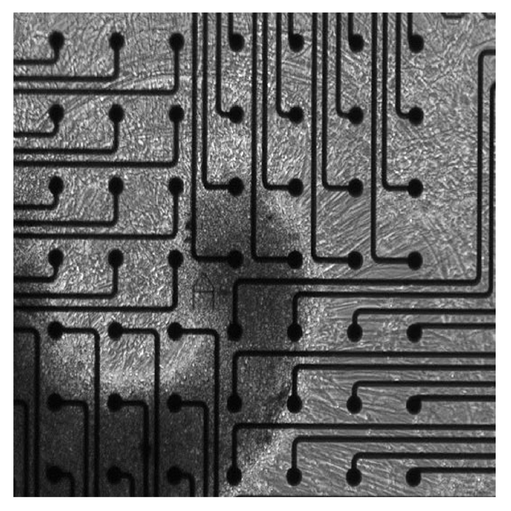
Electrical activity is measured from organoids cultured on electrodes. Cerebral organoids generated from human induced pluripotent stem cells (hiPSCs) exhibit spontaneous neural activity, with increasing complexity as networks mature. Since the microelectrode array is two dimensional, signals recorded reflect the activity of the neurons on the bottom of the organoid. For the best results, ensure contact between the electrodes and the bottom of the organoid.
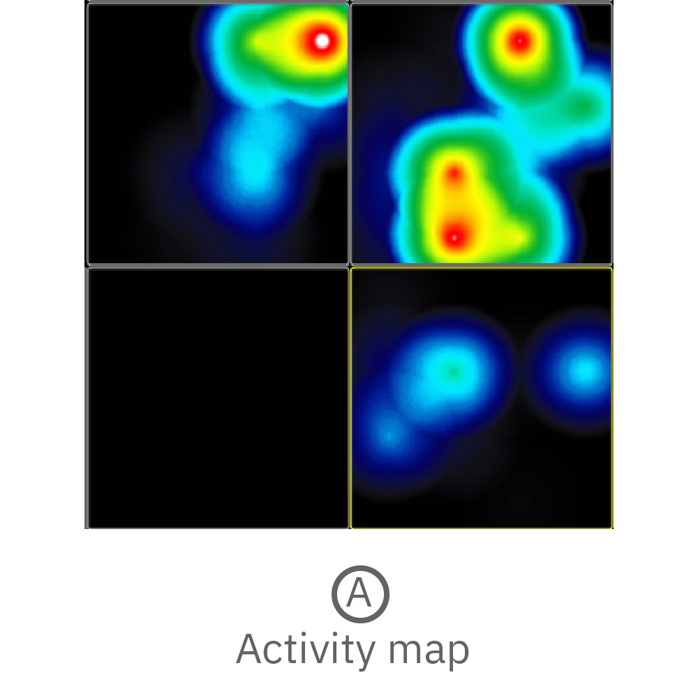
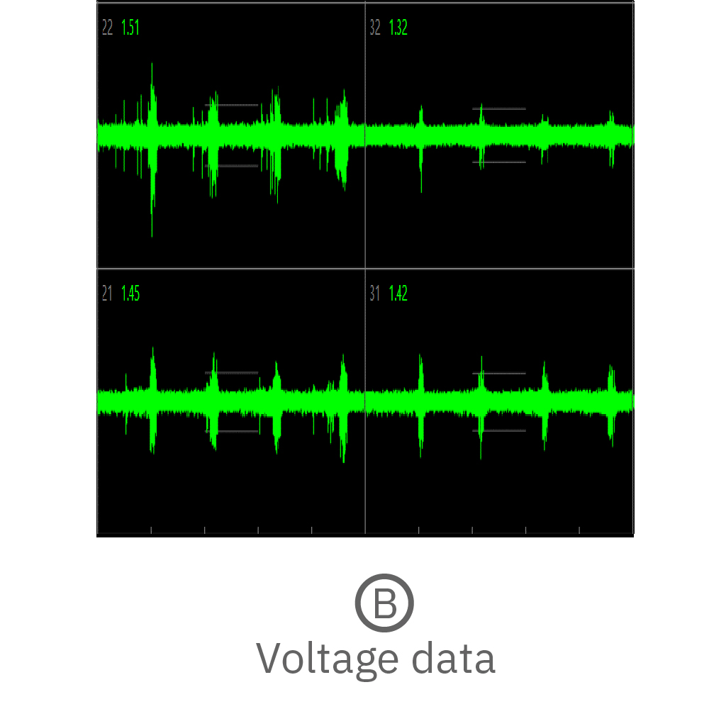
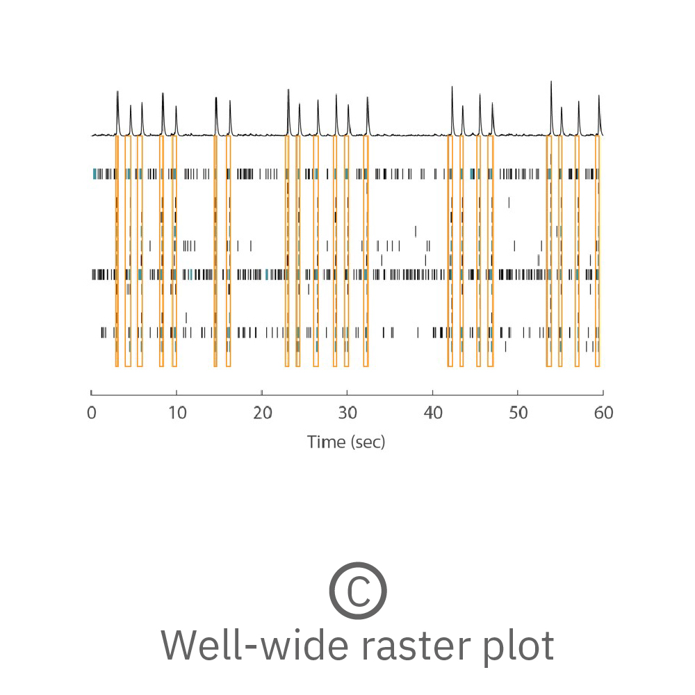
(A) Activity map displaying instantaneous firing rate of four wells with multiple cerebral organoids in each well. (B) Continuous voltage data recorded from four electrodes in one well. Activity is recorded from different sites on the same organoid. (C) Well-wide raster plot showing activity across all 16 electrodes and synchronous network bursting. Teal tick marks indicate electrode bursts, and orange boxes indicate network bursts. Data provided by Maestro customer.
Key findings demonstrated in neural organoid research
Research using the Maestro MEA platform demonstrates how functional electrophysiology can reveal features of organoid biology that cannot be determined from morphology alone.
Examples include:
- Neural spikes and local field potentials can be measured simultaneously from neural organoids.
- LFPs can reveal activity originating beyond regions where surface spiking is detected.
- Cortical organoid network activity can be followed longitudinally over many months as functional complexity develops.
- Cortical organoid maturation has demonstrated electrophysiological features resembling aspects of preterm neonatal brain development.
- Microglia incorporated into cortical organoids can influence the emergence of synchronous and oscillatory network activity.
- Different sites within the same cerebral organoid can display measurable electrical activity and coordinated network bursting.
These studies illustrate how MEA measurements can provide quantitative functional endpoints for developing, comparing, and validating advanced 3D neural models.
Purpose-built MEA solutions for 3D organoid models
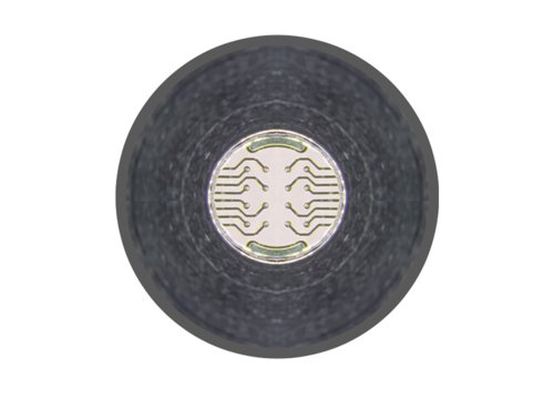
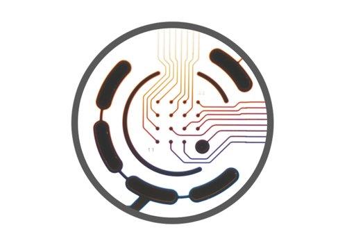
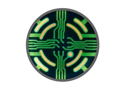
Axion BioSystems offers three MEA technologies designed for 3D organoid and spheroid models. SpheroGuide supports guided placement of larger organoids, SpheroHD provides high-density recording for smaller organoids and spheroids, and 3DMap uses flexible electrodes to interface with three-dimensional tissue surfaces.
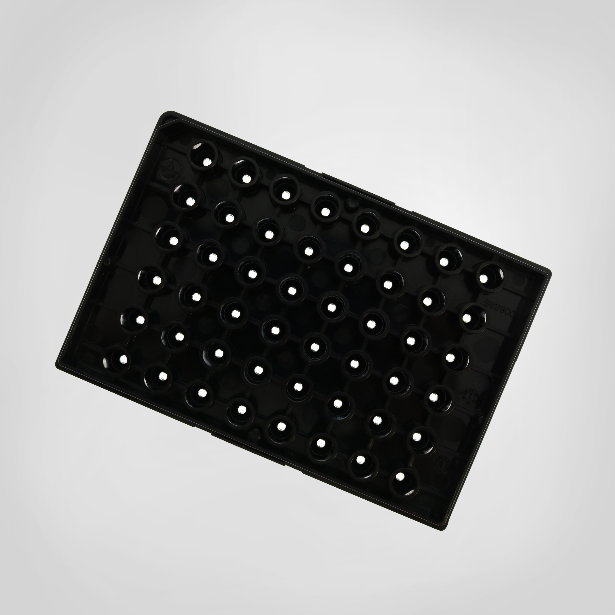
SpheroGuide: guided placement for larger organoids
SpheroGuide MEA plates include an integrated placement funnel that helps position large organoids directly over the electrode array.
The 48-well format enables multiple organoids and experimental conditions to be studied in parallel, with 16 electrodes in each well.
Designed to help with:
- reproducible organoid placement;
- electrode contact;
- multiwell experimental throughput;
- longitudinal recording;
- visual access through a transparent plate bottom.
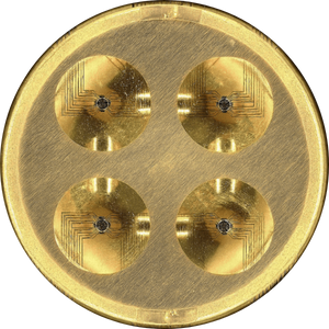
SpheroHD: high-density recording for smaller organoids and spheroids
SpheroHD combines integrated placement features with densely spaced electrodes designed for smaller 3D models.
With 50 µm electrode spacing and multiple microwells, SpheroHD provides a high-density interface for capturing functional activity from compact organoids and spheroids.
Designed to help with:
- small organoid and spheroid placement;
- high-density functional recording;
- improved spatial sampling;
- repeated measurements from the same model.
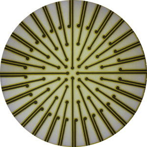
3DMap: flexible electrodes for a three-dimensional interface
3DMap extends MEA recording beyond the conventional flat electrode surface.
Flexible electrode technology is designed to interface with the surface of a 3D organoid, enabling recordings across more of the model while maintaining its three-dimensional form.
Designed to help with:
- interfacing electrodes with a 3D surface;
- increasing access to functional signals around the organoid;
- characterizing complex activity from advanced models;
- preserving organoid morphology during recording.

A complete workflow for neural organoid research
Axion’s live-cell platforms can be used from stem cell culture through mature organoid analysis.
Monitor stem cell cultures: Use live-cell imaging to follow stem cell growth and culture quality before organoid formation.
Track organoid formation and growth: Monitor organoid morphology, size, and development using continuous imaging.
Position the organoid for functional recording: Select an MEA interface designed for the size and geometry of the 3D model.
Record functional neural activity: Measure spikes, bursts, network activity, synchrony, oscillations, and local field potentials.
Follow maturation longitudinally: Return to the same models over time to characterize developmental trajectories or treatment response.
This integrated approach connects morphological and functional measurements throughout organoid development.
Featured resources for neural organoid research
Neural Organoid Publications and Resources
Explore peer-reviewed publications and scientific resources using Maestro MEA for neural organoid electrophysiology.
Explore Neural Organoid Research Resources
Whole Neural Organoid Plating for MEA Recording
Review a step-by-step method for positioning whole neural organoids for spike and local field potential recording.
Neural Organoids on 3DMap MEA Protocol
Neural Spheroids on SpheroHD Protocol
Neural Organoids on SpheroGuide Protocol
Neural Organoid Plating Protocol for MEA Plates
Frequently asked questions about neural organoid MEA assays
Yes. Neural organoids can generate spontaneous extracellular electrical activity that can be recorded with microelectrode arrays.
MEA recordings can quantify neural spikes, bursts, network activity, synchrony, oscillations, and local field potentials.
Microelectrode arrays record extracellular electrical activity generated by neurons within an organoid. MEA recordings can be used to characterize:
- spontaneous neural firing;
- bursting and network bursting;
- network synchrony;
- oscillatory activity;
- local field potentials;
- changes in functional activity during maturation;
- responses to drugs, disease phenotypes, or experimental perturbations.
Because MEA recording is label-free and noninvasive, researchers can return to the same organoid models repeatedly and follow functional development over weeks or months.
Yes. MEA recording is noninvasive and label-free, allowing the same organoids to be measured repeatedly over extended culture periods.
The cortical organoid study highlighted on this page included longitudinal MEA recordings over 10 months.
Three-dimensional organoids present different recording challenges than two-dimensional neural cultures. Specialized designs can help improve organoid placement, electrode contact, spatial sampling, or interaction between electrodes and the 3D model. Axion offers multiple organoid-focused designs for different model sizes and experimental needs.
The appropriate interface depends on the model and experimental goal.
SpheroGuide is designed to help position larger organoids over the electrode area. SpheroHD provides dense electrode spacing and placement features for smaller organoids and spheroids. 3DMap uses flexible electrodes designed to interface with the surface of a 3D organoid.
MEA recordings primarily detect extracellular signals from cells interacting with or near recording electrodes. Local field potential measurements can provide additional information about coordinated population activity.
In the organoid example shown on this page, LFPs were detected in regions where surface spike activity was not observed.
Yes. Because the same models can be recorded repeatedly, MEA is well suited for longitudinal studies of neural organoid development.
Researchers can follow changes in firing, network bursting, synchrony, oscillations, and local field potentials as the model matures.
Yes. MEA measurements can quantify how changes in cellular composition affect neural network function. In research highlighted on this page, transplanted microglia accelerated the emergence of synchronous and oscillatory network activity in cortical organoids.