Traditional neuroscience research has often relied on animal models, but these approaches cannot fully capture the complexity of human brain function. Using human stem cells combined with the Axion BioSystems Maestro MEA platform, Dr. Halliwell’s team developed a human-relevant model to record neural activity, reproduce seizure-like dynamics, and evaluate therapeutic compounds in real time.
In this Coffee Break Webinar, Dr. Robert Halliwell from the University of the Pacific discusses how human cerebral organoids—three-dimensional, stem cell–derived models of the brain—can be used to study epilepsy and discover new anti-seizure medicines.
What you will learn in this 13-minute webinar:
- >> How 3D human cerebral organoids can model neuronal network activity and seizure-like behavior in vitro
- >> How the Axion BioSystems Maestro Edge MEA system enables real-time, long-term recording of brain organoid activity
- >> How anti-seizure compounds can be evaluated for efficacy using a human stem cell–derived brain model
- >> How organoid-based systems are advancing translational neuroscience while reducing reliance on animal models
About the presenter:

Robert Halliwell, PhD
Dr. Robert Halliwell did his graduate training in neuroscience at the University College London and pharmacology at the University of Dundee. He then went on to complete a postdoctoral fellowship in neuroscience at the University of California, Irvine.
Since 2002, at the University of the Pacific, he's taught neuroscience and clinical pharmacology and published work in the history of medicine. His lab addresses the properties of neurons derived from human stem cells for studies in neuropharmacology, neurotoxicology, and disease modeling.
Transcript of the webinar:
Thank you for joining our Coffee Break Webinar.
Today’s topic is Cerebral Organoids to Model Seizures and for the Discovery of Anti-Seizure Medicines.
Our speaker today is Dr. Robert Halliwell from the University of the Pacific. Dr. Halliwell did his graduate training in neuroscience at University College London and pharmacology at the University of Dundee. He then went on to complete a postdoctoral fellowship in neuroscience at the University of California, Irvine.
Since 2002, at the University of the Pacific, he has taught neuroscience and clinical pharmacology and published work in the history of medicine. His lab addresses the properties of neurons derived from human stem cells for studies in neuropharmacology, neurotoxicology, and disease modeling.
Dr. Robert Halliwell:
Thank you for inviting me to present and for the kind introduction. The title of my Coffee Break Webinar today is Cerebral Organoids to Model Seizures and for the Discovery of Anti-Seizure Medicines.
As many people watching this webinar already know, the brain is arguably the most complicated system in the known universe, so finding new medicines for brain disorders is challenging. In addition, studying the living human brain is technically, ethically, and legally very difficult.
To overcome some of these problems, neuroscientists and pharmaceutical scientists have traditionally used animal models to try to understand the brain and to develop new medicines. The problem with this approach is that animals aren’t people, making the search for medicines inefficient, expensive, and, of course, raising questions about the ethics of using animals in experiments as substitutes for humans.
A powerful alternative approach emerged over 20 years ago with the increasing availability of human stem cells to create diverse human tissues. Recognizing this, our lab has been investigating the value of neurons and glia derived from human stem cells to model disease and for drug studies for over 23 years. We have a particular interest in epilepsy.
Epilepsy is a complex neurological disorder with many causes. As my slide says, it is characterized by an occasional, excessive, and disorderly discharge of nerve tissue—a truly accurate description given by the father of neurology, Dr. John Hughlings Jackson, more than 150 years ago.
As can be seen in my slide, epilepsy is the fourth most common neurological disorder, affecting over 65 million people globally. Despite over 32 diverse anti-seizure medicines being available, one-third of patients still suffer seizures. That translates to 15 million people, so there is a pressing need to find new medicines.
The problem with trying to model epilepsy is that it presents in many ways in patients—from dramatic tonic-clonic seizures, when a person loses consciousness, falls, becomes rigid, and convulses, to the patient who loses awareness for a few seconds but doesn’t lose muscle control or fall, as in absence epilepsy.
The common link we can exploit in an in vitro model is that for almost all epileptic seizures, there’s increased brain activity consisting of increased action potential firing, bursts of action potentials, and synchronized abnormal or epileptiform activity in parts of the brain or across the whole brain. This can be measured on the EEG in vivo.
Our first aim in this study was to determine if we could model epileptiform activity in human 3D cerebral organoids.
To make cerebral organoids, we use a human pluripotent stem cell line and a commercial organoid kit. We currently maintain and mature the cerebral organoids for up to eight months. Interestingly, in 2D culture, we can maintain these neurons and glia for up to two years, and we’ve published both methods for reference.
This is an oblique image of part of the surface of a fixed organoid at 130 days in vitro (about four and a half months), immunolabeled with beta III-tubulin identifying neurons in green, GFAP indicating glial cells in red, and DAPI for nuclear DNA shown in blue. Even in this relatively simple image, it can be seen that these brain organoids are complex and even form ventricle-like structures.
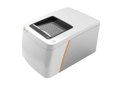
For the next step, we determined the electrophysiological activity of our brain organoids by exploiting the power of multi-electrode arrays, or MEAs, to record from hundreds of cells over many weeks and months to establish their functional maturation.
We use the MEA Maestro Edge system, transferring the organoids at 40 days in vitro into six-well MEA plates, with each well containing 64 electrodes, giving us a total of 384 electrodes per plate.
Recording the baseline activity of neurons in the organoids is crucial before any investigation of provoking abnormal or epileptiform activity can be conducted.
Here is an MEA recording from a brain organoid at 194 days in vitro (about six and a half months). We’re looking at the view from the AxIS software display, showing voltage traces from the 64 electrodes in one well. The green box in the top left corner indicates that we’re looking at activity in well five. In the timeframe of about 40 seconds, 40 of the 64 electrodes detect action potentials from neurons sitting above these electrodes.
The lower part of the screen is a raster display in real time, with every white dot representing an action potential. The red dots represent a burst of action potentials from a single cell, and a line of red dots indicates a network burst of action potentials. This spontaneous neural activity is typical of our cerebral organoids at this stage of their development.
This figure summarizes the electrophysiological maturation of neural organoids over eight months, with an image in the left corner of an organoid at seven weeks on the MEA. At the top of the figure are examples of recordings of action potentials at weeks 8, 20, and 30, with a snapshot below each trace of a heat map of active electrodes in a well.
Note that more electrodes detect spiking and cells fire at higher frequencies as the organoid matures. The plots below quantify the spike rate, bursts, and synchronized firing from week 7 to week 30. All these measures increase up to week 26 because stem cells are differentiating and maturing into neurons. Glial cells are also proliferating from about four months, and synaptogenesis rapidly increases from four to eight months.
These neural developments all underlie the enormous increases in burst firing and synchronized activity and are consistent with observations in vivo, indicating that organoid maturation is under genetic control.
The next step in the study was to determine the neurochemical basis of activity in our brain organoids. To do this, we tested the impact of drugs that act at specific synaptic receptors and ion channels on the spontaneous spike rate in the organoids.
As can be seen in this histogram summary, the sodium channel blocker TTX reduced the action potential rate, whereas the potassium channel blocker XE991 increased it. These data confirm that we have sodium- and potassium-ion-channel-dependent action potentials.
The summary also shows that the GABA receptor antagonists bicuculline and picrotoxin increased spike frequency, whereas the glutamate receptor antagonist kynurenic acid reduced spike activity.
Together, these data show that our brain organoids have formed inhibitory and excitatory neural circuits and that synaptic neurotransmission occurs through GABA_A and ionotropic glutamate receptors, respectively.
In addition, we also see that three pharmacologically distinct anti-seizure medicines—carbamazepine, ethosuximide, and diazepam—modulate ongoing spontaneous neurophysiological activity consistent with in vivo observations.
Our next task was to determine if we could evoke epileptiform activity in the brain organoids. The potassium channel blocker 4-aminopyridine is a powerful convulsant in animals and in cultured cells.
As can be seen in this slide, when we applied 4-aminopyridine to the brain organoids, it evoked an enormous increase in all measures of neural activity paralleling epileptiform activity in vivo. The left half of the figure shows an MEA recording of action potentials at baseline and then in the presence of 4-AP. Beneath is a snapshot of the heat map of active electrodes in a well in each of these two conditions.
The histograms on the right side of the slide confirm that spike rates, cell bursts, network bursts, and synchronized firing were all significantly increased by 4-aminopyridine, consistent with epileptiform activity in vivo.
Our next question was whether anti-seizure medicines could reduce or inhibit epileptiform activity in these brain organoids.
As shown in these four histogram summaries, 4-aminopyridine evoked increases in spiking, cell bursts, network bursts, and synchronized firing across the organoid as described in the previous slide. In addition, the figure shows that the anticonvulsant diazepam, a GABA receptor potentiator, reduced or completely inhibited these measures of epileptiform activity.
Notably, additional experiments in our lab, not included in this webinar, have also shown that other anti-seizure medicines—including ethosuximide, a T-type calcium channel blocker used for absence epilepsy, and carbamazepine, a sodium channel blocker used for partial and generalized epilepsy—can also significantly inhibit epileptiform activity.
These data indicate the potential of human cerebral organoids in anti-seizure medicine discovery.
Our conclusions from this study are:
- First, human cerebral organoids form 3D circuits composed of neurons and glial cells.
- Second, they generate spontaneous neurophysiological activity, which increases in both volume and complexity as the organoids develop and mature.
- Third, from our extensive pharmacological experiments, we can conclude that neurons in these brain organoids express diverse functional receptors and ion channels that underlie action potential generation and synaptic neurotransmission.
- Fourth, powerful inhibitory and excitatory circuits are formed, and synaptic neurotransmission occurs through GABA_A and ionotropic glutamate receptors.
- Fifth, convulsant drugs can evoke hyperactivity in cerebral organoids that parallel EEG patterns seen in epilepsy.
- And finally, epileptiform activity can be inhibited by diverse anti-seizure medicines, making human 3D brain organoids combined with MEA technology powerful tools in drug discovery for neurological disorders.
To finish, I would like to acknowledge the support for our research provided by the Thomas J. Long School of Pharmacy at the University of the Pacific here in sunny Stockton, California.
For anyone looking for more information on our work, the links to the three papers below will provide a lot more detail.
Thank you for listening. That concludes today’s Coffee Break Webinar. If you have any questions regarding the research presented, or if you’re interested in presenting your own research with microelectrode array technology or impedance-based assays, please forward them to coffeebreak@axionbio.com.
Thank you for joining today’s Coffee Break Webinar. We look forward to seeing you again.