Biology, like you’ve never seen before
Live-cell imaging captures dynamic, time-dependent cellular processes capable of adding new dimensions to your research. At Axion BioSystems, we offer innovative tools to expand the capabilities of your lab and accelerate discovery and therapeutic development. See how we are advancing research with our live-cell imaging platforms.

350+
systems sold

200,000+
Omni scans

300+
peer-reviewed papers
Why live-cell imaging?
Live-cell imaging has revolutionized cell biology research, offering distinct advantages compared to traditional endpoint assays and other conventional cell analysis methods.

Complete, real-time datasets
Measure kinetics and gather insights that endpoint assays miss

Physiologically relevant assays
Assay living cells in optimal conditions with environmental control

Experimental flexibility
Record from nearly any culture vessel, multiplex readouts, and adapt workflows

No complicated steps
Reduce hands-on time and save money with a simple assay
Dynamic, real-time cellular processes >>
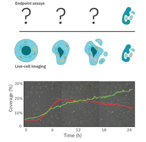
Biologically relevant results
Biology changes over time. Capture the entire process in real time and never miss a critical event during your experiments.
In this example, early and late apoptosis are simultaneously tracked with pSIVA (green) and propidium iodide (red). Real-time data reveals the dynamic interplay throughout the experiment.
Across the entire well >>
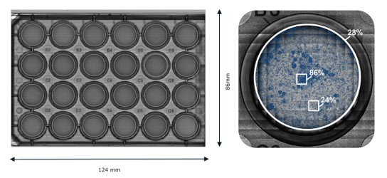
Fast, AI-driven analysis
With fast, whole-well brightfield imaging capabilities, reduce variability by removing sampling bias. Create high resolution, full-vessel scans compatible with a wide array of culture formats.
Optimizing CAR structures to enhance efficacy in solid tumours
Learn how researchers used real-time assays on the Omni to evaluate potency and optimize CAR structures.
Watch more
Easy, fast analysis with intuitive software >>
Our software modules provide the tools for fast, accurate analysis and the flexibility to customize the platform to suit your research needs. Monitor growth and death in monolayers, colonies, or organoids with whole-well brightfield imaging, or multiplex with fluorescence assays to examine specific mechanisms or markers. The examples below highlight some of the capabilities of the Omni platform.
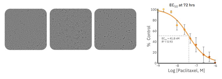
Confluency Module: Whole-well growth and cytotoxicity
Changes in confluence can indicate alterations in cell proliferation, migration, or death. Assess the health and viability across the entire plate in real time. In this example, real-time cytotoxicity was evaluated in monolayers with an anticancer agent. Read more
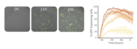
Fluorescence Module: Versatile assays
Red and green fluorescence channels add compatibility with a wide range of assays. Multiplex your experiments with live-cell compatible labels or with one of many traditional endpoint assays. In this example, GFP was used to optimize transduction protocols using BacMam.
Read more
Kinetic live-cell cancer assays >>
Monitor the survival, growth, and mobility of cancer cells over time for deep inisghts into cellular behavior and therapeutic response. The Clonogenic Assay Module and Scratch Assay Module streamline the analysis of these common oncology assays to advance our knowledge of cancer biology and facilitate the development of more effective anticancer strategies.
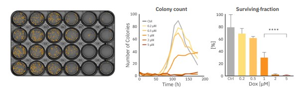
Clonogenic Assay Module: Fast, accurate colony counts
For accurate counting in low-density clonogenic assays, automated whole-well imaging is critical. Clonogenic assays have a strong time-dependence as colonies grow and merge over time. Real-time imaging ensures your analysis can be done at the optimal time. In this example, cancer cells were dosed with increasing concentrations of an anticancer agent. Colonies were quantified automatically across the entire plate over 170 hours to calculate the dose response. Read more
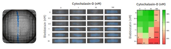
Scratch Assay Module: Dynamic cell migration
Cell migration and wound healing are dynamic processes that can’t be captured in a single endpoint. A scratch assay tracks the closure of a controlled gap in a confluent cell culture and is common in oncological and regenerative medicine research. In this example, blebbistatin and cytochalasin-D alter cell motility and inhibit gap closure. By quantifying the entire scratch area, variability introduced by manual scratch placement can be minimized. Read more
Culture quality monitoring >>
A consistent differentiated cell begins with consistent starting material. Noninvasively track cellular processes crucial to development and differentiation. Improve your process with real-time visualization and continuous monitoring of stem cell behavior, organoid formation, and response to environmental cues.
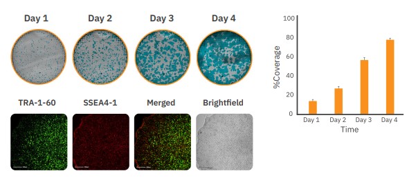
iPSC Module: Monitor growth and quality
Track the growth of your pluripotent stem cell cultures label-free to ensure you always passage at the right time. Coverage of stem cell colonies was monitored so cells could be passaged when they reached 70%. Pluripotent markers were confirmed at day 4 through fluorescent staining using the Fluorescent Module. Read more
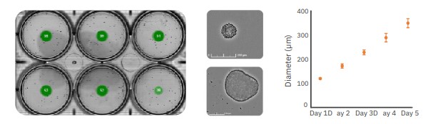
Organoid Analysis Module: Track formation and growth
The quantity, size, and roundness of embryoid bodies (EBs) were tracked to ensure normal, consistent formation and growth. Normal handling and maintenance often moves EBs, organoids, and spheroids in the dish, making single-site imaging unsuitable for this purpose.
Read more
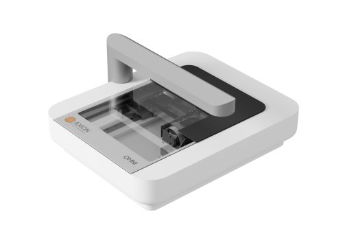
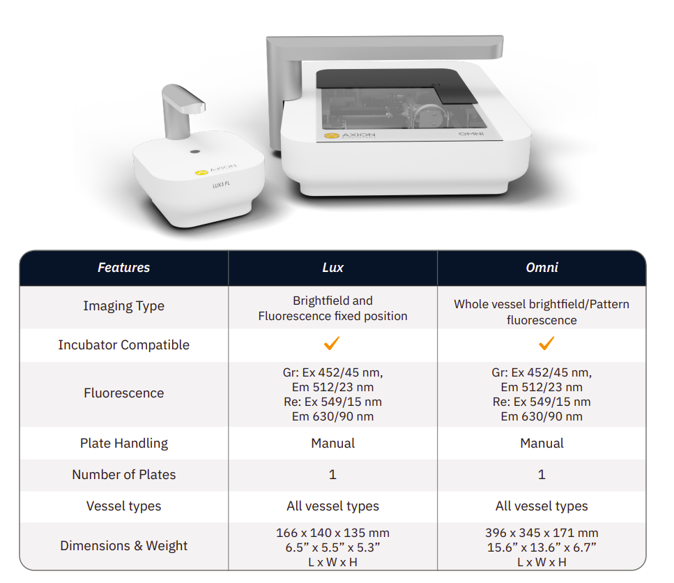
Live-cell imaging from your incubator >>
Whole-well brightfield or fluorescence imaging with single or multiplate options and a range of easy-to-use software modules lets you customize your analysis.

Whole-plate imaging

Incubator-based design

Flexible workflows
Our commitment to our customers
With over 15 years of experience bringing innovative new products to our customers, we strive to accelerate your research by making live, functional biology more accessible. Our design philosophy is to ensure all of our products are:

Flexible
Hardware designed for broad, integrated functionality in one instrument

Easy to use
Intuitive instruments, consumables, and software for fast, easy adoption

Smart technology
Easy to run with no complicated steps, saves time and money.