This protocol details the use of impedance, via the MEA Viability Module, to monitor cell attachment on CytoView MEA Plates. Impedance measurements detect the presence of intact cell membranes on the same recording electrodes used to monitor functional activity of neurons and cardiomyocytes. As such, the MEA Viability Module can be used to track changes in cell coverage over time, implement quality control criteria, or evaluate the structural toxicity of drugs or chemicals. This protocol requires a CytoView MEA Plate, a Maestro Pro or Edge, and the MEA Viability Module.
Cell Culture
- Seed and culture the cells on a CytoView MEA Plate according to cell supplier recommendations. Note: CytoView MEA plates are required for MEA Viability measurements.
Perform a MEA Viability Measurement
- Perform a MEA Viability measurement each time you measure the electrophysiological activity of the cells using a Viability configuration. Tip: The viability data and electrical activity data are stored in the same RAW file for easy access and analysis with Axion software tools.
- Place the CytoView MEA Plate in the Maestro Pro or Edge, and switch to the “Configuration” → “Neural + Viability Real-Time” → “Spontaneous + Viability” configuration or the “Configuration” → “Cardiac + Viability Real-Time” → “Field Potential + Viability” configuration. Tip: Viability measurements are reported in units of “Resistance (㏀)”.
- Press Record. The MEA Viability measurement will be acquired first, followed by offset correction and the corresponding Neural or Cardiac measurement. Press Record again (or Stop) to end the measurement. Tip: Viability measurements are compatible with scheduled recording to collect measurements over time. Note: The MEA Viability measurement takes 1 minute to complete. After the MEA Viability measurement is acquired, you may view the data in the Activity Map using the source selector drop-down menu.
- Repeat steps 3-4 throughout the maturation of the culture, or, for a dosing experiment, before and after the perturbation. Note: The absolute value of the MEA Viability measurement indicates cell coverage. However, it is best used as a relative measure to assess changes in cell viability. Tip: The MEA Viability measurement includes the Resistance of the electrode itself (~8㏀). For this reason, control compounds may be helpful to establish the range for covered vs. uncovered electrodes.
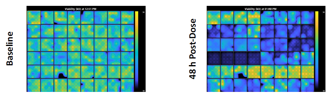
hiPSC-derived neurons (NeuCyte Labs) were seeded on a CytoView MEA 48-well plate and cultured for 14 days. The MEA Viability module was used to monitor cell attachment before (Baseline, left) and after (Post-Dose, right) the addition of neurotoxic compounds. The impedance measurements were acquired at 0.5, 4, 24, 48, and 72 hours post-dose.
Analyze Viability Data in AxIS Navigator
- Load a RAW file into AxIS Navigator using the “File → Open Recording” menu, or by dragging the RAW file into the stream tree.
- Right click to apply the appropriate configuration (e.g. Spontaneous + Viability).
- Press Record to begin the analysis, producing a Statistics Compiler CSV file.
- Repeat steps 6-8 for each recording you intend to analyze. Note: Batch processing may be used to process multiple MEA Viability files with the same configuration. Additional analysis may be performed with the Neural Metric Tool (NMT) or Cardiac Analysis Tool (CAT).
Create Viability Data Figures with the AxIS Metric Plotting Tool
- Press the “Baseline” button in the AxIS Metric Plotting Tool to load a Statistics Compiler CSV file.
- Press the “Comparison” button to select any additional Statistics Compiler CSV files for analysis.
- Select “Resistance - Avg (㏀)” from the Metric drop-down menu. Note: Using a control compound, like Triton X-100, that lyses the cell membrane can be helpful when analyzing MEA Viability measurements in a dosing experiment. For further details, see the MEA Viability FAQ in the AxIS Navigator User Guide.
- Repeat steps 10-12 for each experiment.
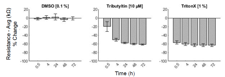
The Vehicle control (0.1% DMSO) had no effect on viability as quantified through resistance measurements. Triton X-100 (1%) lysed the cell membranes, serving as a positive control. Tributyltin (10 𝜇M) gradually killed the hiPSC-derived neurons over the first 24 hours.
Required Materials
| Item | Vendor |
|---|---|
| CytoView MEA Plate | Axion BioSystems |
| Maestro Pro or Edge | Axion BioSystems |
| AxIS Navigator (version 3.4 or higher) | Axion BioSystems |
| MEA Viability Module | Axion BioSystems |
| AxIS Metric Plotting Tool (version 2.4.4 or higher) | Axion BioSystems |
| Surface Coating (Fibronectin, PEI, Laminin, etc) | Various |
| Cell Culture Media | Various |