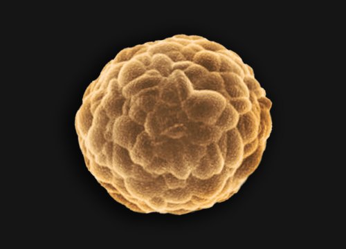
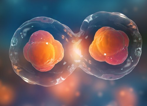
iPSC Organoid Models
Explore advanced human-relevant models with iPSC organoids
Learn More→
AI-powered imaging analysis uses trained image analysis models to identify, segment, track, and quantify biological features from live-cell imaging data. For researchers working with complex cultures, stem cells, colonies, or organoids, this helps turn large image datasets into interpretable results with less manual analysis.
With Axion BioSystems’ Omni live-cell imaging systems and Axion Portal software, researchers can monitor cells over time, analyze whole-well images, and generate quantitative readouts using AI-enabled analysis modules designed for common live-cell workflows.
Live-cell imaging can capture rich biological information, but the analysis can quickly become the limiting step. A single experiment may generate thousands of images across wells, time points, and conditions. Manually reviewing those images can be slow, subjective, and difficult to reproduce across users.
AI-enabled analysis helps researchers move from image collection to biological interpretation faster. Instead of spending time adjusting segmentation settings or counting objects by hand, researchers can use trained analysis modules to measure growth, confluence, colony formation, iPSC culture health, and organoid development across the full experiment.
The Omni is a great device for following organoid growth and morphogensis over time accross multiple conditions. The ability to image entire plates in the incubator allowed us to capture unique events and generate large data sets at ease.
AI imaging analysis with Omni and Axion Portal fits into standard live-cell workflows. Researchers culture their cells normally, image them over time, and use application-specific analysis modules to generate quantitative results.

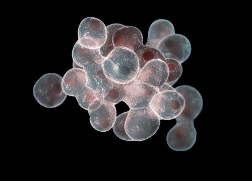
Cells, colonies, iPSCs, or organoids are cultured in compatible vessels and imaged using the Omni live-cell imaging system. The system captures detailed whole-well images over time, allowing researchers to monitor the same cultures longitudinally.

Researchers choose the module that matches the biology and assay goal, such as Confluency, Organoid Analysis, iPSC, or Colony Analysis.

The software identifies relevant biological features and generates quantitative measurements. This reduces the need for manual counting, repeated threshold adjustment, or user-by-user segmentation decisions.

Researchers can review whole-well images, time-lapse data, and analysis plots to understand how cultures change over time. Results can be compared across wells, conditions, and time points.

The final step is the most important one: using the data to answer the experimental question. Did the cells proliferate? Did treatment alter growth? Did organoids change size or morphology? Were iPSC cultures ready for the next step?
AI helps make the analysis faster and more consistent, so researchers can focus on what the results mean.
Axion’s AI analysis tools are developed with input from software engineers and application scientists. The goal is to build models that recognize the biology researchers care about, not just pixels on a screen.
Omni captures the full well, helping researchers see heterogeneity that may be missed with limited fields of view. This is especially important for organoids, colonies, and unevenly distributed cultures.
Axion Portal uses cloud-based tools to support fast processing, data access, collaboration, and software updates. The Portal page highlights cloud access from any device, automated user alerts, data organization, collaboration, and automatic updates.
AI analysis is most useful when it makes the workflow simpler. Axion’s imaging analysis modules are designed to help researchers move from image acquisition to quantitative results with fewer manual steps.

Axion Portal includes analysis modules for a range of live-cell imaging workflows, including Confluency, Organoid Analysis, iPSC monitoring, Colony / Clonogenic Analysis, Fluorescence, and Scratch Assay workflows.
Manual analysis can be slow, subjective, and difficult to reproduce across users. AI-enabled analysis helps reduce repetitive manual steps and supports more consistent quantification across wells, plates, and experiments.
Yes. Omni systems are designed for live-cell imaging over time, allowing researchers to monitor the same cultures across multiple time points. This is useful for growth, differentiation, treatment response, organoid development, and colony formation studies.
No. AI helps process and quantify the image data. The scientist still reviews the results, evaluates assay quality, and interprets the biology.
Whole-well imaging gives a more complete view of the culture. This can be important when cells, colonies, or organoids are unevenly distributed or when rare events occur outside a small field of view.
No. Axion Portal provides application-specific modules so researchers can analyze common live-cell imaging workflows without developing custom pipelines.