Add Action Potential Insight to a Scalable Cardiac MEA Workflow with LEAP
On-demand webinar
Cardiac electrophysiology assays often require researchers to balance mechanistic detail with experimental scale. Patch-clamp recordings provide direct access to individual action potential waveforms, while microelectrode array assays enable scalable, label-free measurements from functional cardiomyocyte monolayers.
In this on-demand webinar, Vishwanath Lanka, Field Application Scientist at Axion BioSystems, explains how local extracellular action potential technology, or LEAP™, brings action-potential-like waveform information into the familiar cardiac microelectrode array workflow.
Watch the webinar to learn how LEAP adds waveform depth without giving up MEA scale.
What is LEAP?
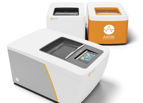
LEAP is an extracellular recording capability available on the Maestro MEA platform. During LEAP induction, coupling between the cardiomyocyte syncytium and selected electrodes is enhanced. This changes the recorded signal from a conventional field-potential waveform into a larger waveform that more closely resembles a cardiac action potential.
The resulting signal includes distinct depolarization, plateau, and repolarization phases, allowing researchers to evaluate action potential morphology from intact cardiomyocyte monolayers.
Because LEAP uses the same cells, MEA plates, and recording platform as a standard cardiac MEA experiment, researchers can add action-potential-like information without transferring their cultures to a separate assay or instrument.
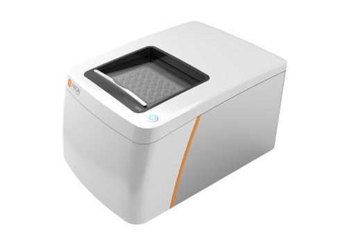
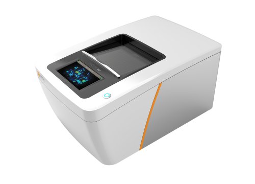
Explore how the Maestro MEA platform supports scalable, label-free electrophysiology experiments.
What will you learn in this webinar?
In this presentation, you will learn:
- How LEAP bridges the gap between patch clamp detail and MEA scalability
- Why action potential morphology matters in cardiac electrophysiology studies
- Tips and best practices for performing LEAP assays
- How LEAP-derived metrics reveal changes in depolarization, plateau, and repolarization behavior
- Example pharmacology data for compounds affecting calcium, potassium, and hERG channel activity
Researchers new to extracellular electrophysiology can also review the fundamentals of microelectrode array technology, including how MEAs record electrical activity from cardiomyocyte cultures.
Measuring cardiac action potential morphology
Action potential morphology can provide insight into the ion-channel mechanisms underlying cardiomyocyte function.
The depolarization phase is predominantly influenced by sodium currents, the plateau phase is strongly influenced by calcium currents, and repolarization is primarily governed by potassium currents. Compounds that affect these channels may therefore produce changes in different regions of the action potential waveform.
LEAP enables researchers to quantify morphology-based endpoints such as:
- Rise time for evaluating depolarization-related effects
- APD30, APD50, and APD90 for measuring action potential duration at different stages of the waveform
- Triangulation for evaluating changes in repolarization shape
- Early afterdepolarization detection for identifying potentially arrhythmic waveform behavior
These measurements complement conventional cardiac field-potential analysis and provide an additional view of compound-induced electrophysiological changes. Explore the broader capabilities of cardiac MEA technology.
Pharmacology examples presented in the webinar
The webinar demonstrates how LEAP captures expected changes in action potential morphology following treatment with compounds that affect different cardiac ion channels.
Nifedipine and calcium-channel inhibition
Nifedipine, an L-type calcium-channel blocker, produces dose-dependent shortening of the LEAP waveform. This response is reflected in reductions in APD30, APD50, and APD90, demonstrating how LEAP can quantify changes associated with the plateau phase of the cardiac action potential.
E-4031 and hERG-channel inhibition
E-4031 blocks hERG potassium channels involved in cardiac repolarization. In the presented data, E-4031 prolongs the LEAP waveform, with higher concentrations also producing early afterdepolarizations.
The action-potential-like LEAP signal makes these repolarization changes and arrhythmic events easier to identify and quantify than they may be in a conventional field-potential waveform.
Tolterodine and terodiline
The comparison between tolterodine and terodiline demonstrates why action potential shape can provide information beyond action potential duration alone.
Although both compounds prolong the action potential, terodiline produces more pronounced triangulation of repolarization. LEAP captures this distinction, illustrating how waveform morphology may help differentiate electrophysiological responses that appear similar when evaluated only by duration.
These applications are especially relevant for researchers investigating compound effects and potential proarrhythmic liabilities. Discover additional approaches for evaluating comprehensive in vitro cardiac activity.
Incorporating LEAP into a cardiac MEA experiment
LEAP is designed to complement an established cardiac MEA assay rather than replace it.
Researchers can induce LEAP on selected electrodes to collect baseline and post-dose recordings, or complete a standard field-potential workflow before adding LEAP at the end of the experiment. Flexible electrode selection allows part of the array to remain available for conventional field-potential measurements.
The webinar also discusses practical considerations for obtaining high-quality recordings, including:
- Selecting an appropriate MEA plate and well format
- Beginning with healthy cardiomyocyte cultures and consistent baseline field potentials
- Optimizing coating, plating density, maturation time, and media conditions
- Planning electrode use when multiple LEAP recordings are required
- Matching analysis endpoints to the expected biology
- Combining LEAP with electrical or optical stimulation when evaluating pacing or rate-dependent effects
The Cardiac Software Module supports action-potential, field-potential, propagation, and contractility measurements within the Maestro MEA workflow.
A more complete view of cardiac function
LEAP is one component of Axion BioSystems’ broader cardiac assay portfolio. On the Maestro MEA platform, researchers can examine multiple aspects of cardiomyocyte function, including:
- Action potential morphology
- Extracellular field potentials
- Electrical propagation and conduction
- Mechanical contractility
Combining these complementary measurements can provide a more complete view of cardiomyocyte physiology from early discovery through cardiac safety assessment.
Learn how label-free cardiac contractility measurements can complement electrophysiology data by evaluating contraction amplitude, timing, and excitation-contraction coupling.
Watch the webinar
Discover how LEAP adds detailed action potential morphology to a scalable, label-free cardiac MEA workflow.
Presenter:
Vishwanath Lanka
Field Application Scientist
Axion BioSystems
Contact an Axion BioSystems expert to discuss how LEAP and Maestro MEA assays could support your cardiac research or drug discovery workflow.
For research use only. Not for use in diagnostic procedures.