In vitro microelectrode array (MEA) technology is a sophisticated method used to measure and analyze the electrical activity of cells in a controlled laboratory setting.
Microelectrode arrays measure the field potential, or activity, across an entire population of cells, detecting patterns that would elude single cell techniques such as patch clamp electrophysiology while providing more detail and resolution than techniques like voltage sensitive dyes.
Available in a range of formats, MEA systems are the perfect solution for high-throughput, detailed, in vitro electrophysiology.
Topics:
>> What is microelectrode array?
>> Microelectrode array advantages
>> How does microelectrode array work?
>> Key components of an in vitro MEA assay
>> Applications of microelectrode array electrophysiology
>> Neural electrophysiology on microelectrode arrays
>> Cardiac electrophysiology on microelectrode arrays
>> Microelectrode array systems
>> Microelectrode array plates
>> Frequently asked questions about microelectrode array systems
What is microelectrode array?
A microelectrode array is a grid of tightly spaced microscopic electrodes that can detect the electrical signals of cells in close proximity. Microelectrode arrays can be embedded in the bottom of a well and cells can be cultured over the electrodes for passive, noninvasive monitoring of electrical activity. These signals are vital for understanding cell communication, network activity, and the physiological behavior of tissues. An in vitro MEA system allows researchers to study the electrical properties of cells in a way that mimics the behavior of the cells in living organisms, providing valuable insights for fields such as neuroscience, pharmacology, toxicology, and disease modeling.
Watch the full video to discover if an MEA assay is right for your research.
Microelectrode array advantages
Microelectrode array assays have become an indispensable tool in labs worldwide. Detailed electrophysiological characterization is critical for understanding the mechanisms of disease and the safety and efficacy of therapeutics. Why have labs adopted MEA technology? An MEA assay offers a powerful combination of throughput, resolution, and ease of use.

Detailed functional data
Dynamic spatial and temporal functional data reveals how cells fire and interact in culture

Noninvasive, real-time monitoring
Noninvasive electrodes track activity over time for maximum experimental flexibility

Easy assay technique
Requires only basic culture techniques with no labels, dyes, or complicated steps

High-throughput capacity
Simultaneous recording of electrical activity from 6 up to 96 wells of MEA data
How do microelectrode arrays work?
Electrically active cells such as neurons or cardiomyocytes produce an extracellular field potential. Microelectrode array (MEA) plates have a grid of tightly spaced electrodes embedded in the culture surface of each well. Cells can be cultured over the electrodes, allowing for the measurement of their extracellular field potential. Each electrode captures the activity on a microsecond timescale providing both temporally and spatially precise data, resulting in a detailed electrophysiological profile.
Microelectrode arrays are compatible with many types of electrically active cells such as cardiomyocytes, neurons, retinal cells, and muscle cells.
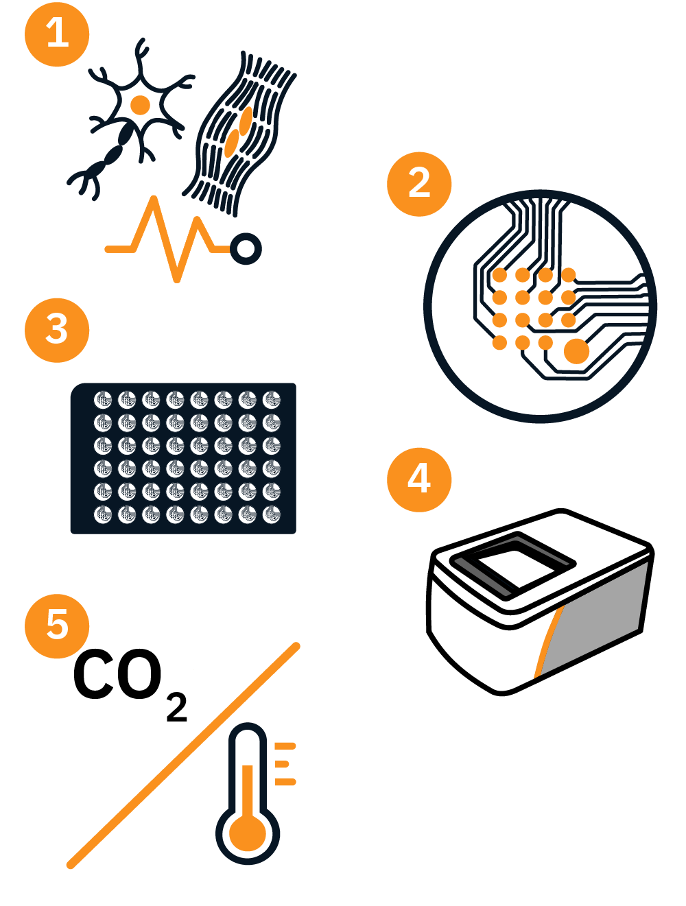
Key components of an in vitro MEA assay
- 1. Electrically active cell culture: In vitro MEA systems support a variety of action potential-firing cell types and are compatible with primary and stem cell-derived cells in 2D culture, 3D organoid or spheroid culture, or slices.
- 2. Microelectrodes: The electrodes on the MEA plate are responsible for detecting the electrical activity generated by cells. MEA electrodes can be made in a variety of sizes, shapes, spacing, and biocompatible materials, depending on the purpose.
- 3. MEA plate: The electrodes are embedded in the culture surface of an MEA plate, which can range from single well up to 96-well formats, ideal for in vitro MEA assays.
- 4. MEA system: The MEA system is crucial for signal amplification, filtering, and digitization. Depending on the MEA system used, it may have additional capabilities like stimulation, multimodal acquisition (action potential contractility, viability), or environmental control.
- 5. Environmental control: Since cell cultures need to be maintained in a controlled environment (temperature, humidity, CO2 levels), MEA systems should have a quality environmental control. This allows researchers to monitor cell behavior over long periods under conditions that simulate those found in living organisms.
(1) Electrically active cells such as neurons or cardiomyocytes produce an extra-cellular field potential.
(2) Axion’s microelectrode array (MEA) plates have a grid of tightly spaced electrodes embedded in the culture surface of each well.
(3) Cells can be cultured over the electrodes, allowing for the measurement of their extracellular field potential.
(4) Each electrode captures the activity on a microsecond timescale providing both temporally and spatially precise data, resulting in a detailed electrophysiological profile.
Applications of microelectrode array electrophysiology
In vitro MEA systems are powerful tools for studying the electrical activity of cells, providing insights into disease mechanisms, and drug responses. The ability of microelectrode arrays to record noninvasively, label-free, and in real time in high-throughput well formats gives them an advantage for monitoring many conditions over short or long periods of time. As such, microelectrode arrays are commonly found in labs conducting disease modeling, drug screening, toxicology testing, and stem cell research. While compatible with many types of cells, microelectrode arrays are most commonly used in neural and cardiac research.
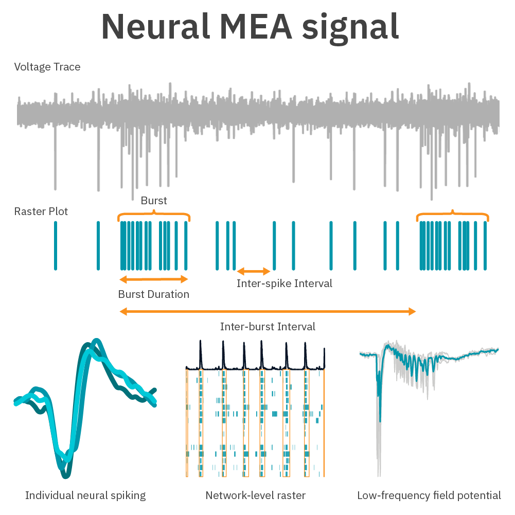
An example of neural signals and metrics measured by microelectrode arrays. A microelectrode array can measure individual neuronal spikes from one electrode, spiking across multiple electrodes, and low-frequency, EEG-like field potentials. Spike timing and clustering can be quantified to characterize neural network patterns, as displayed by a raster plot.
Neural electrophysiology on microelectrode arrays
When neurons are cultured on top of an MEA, they connect synaptically to form a network. There are many types of neurons that fire and communicate in many distinct ways. Using Maestro microelectrode array (MEA) technology, any scientist can quickly and easily measure neural network behavior.
Applications include, but are not limited to:
- >> neural characterization and development
- >> neural co-cultures
- >> neural optical and electrical stimulation
- >> neural organoids/mini-brains
- >> neurological diseases such as Alzheimer's or epilepsy
- >> neuromuscular junctions
- >> neurotoxicity and safety
- >> pain
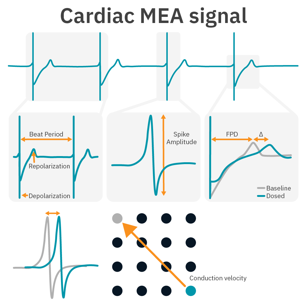
An example of cardiac signals and metrics measured by microelectrode arrays. A microelectrode array measures similarly to an EKG, with a clear depolarization and repolarization. The time difference of depolarization across the array can be used to track conduction across the microelectrode array.
Cardiac electrophysiology on microelectrode arrays
When cardiomyocytes are cultured on top of an MEA, they attach and connect to form a spontaneously beating sheet of cells, called a syncytium. When one cardiomyocyte fires an action potential, the electrical activity propagates across the syncytium causing each cell to fire and then contract. The electrodes detect each individual action potential and contraction, as well as the propagation of this activity across the array. The propagating electrical signal is detected by the electrodes as an extracellular field potential.
Applications include, but are not limited to:
- >> cardiac characterization
- >> cardiac differentiation and maturation
- >> cardiomyocyte pacing using optical or electrical stimulation
- >> evaluation of cardiotoxicity and proarrhythmic compounds
- >> cardiomyocyte inotropy and excitation-contraction coupling
Microelectrode array systems
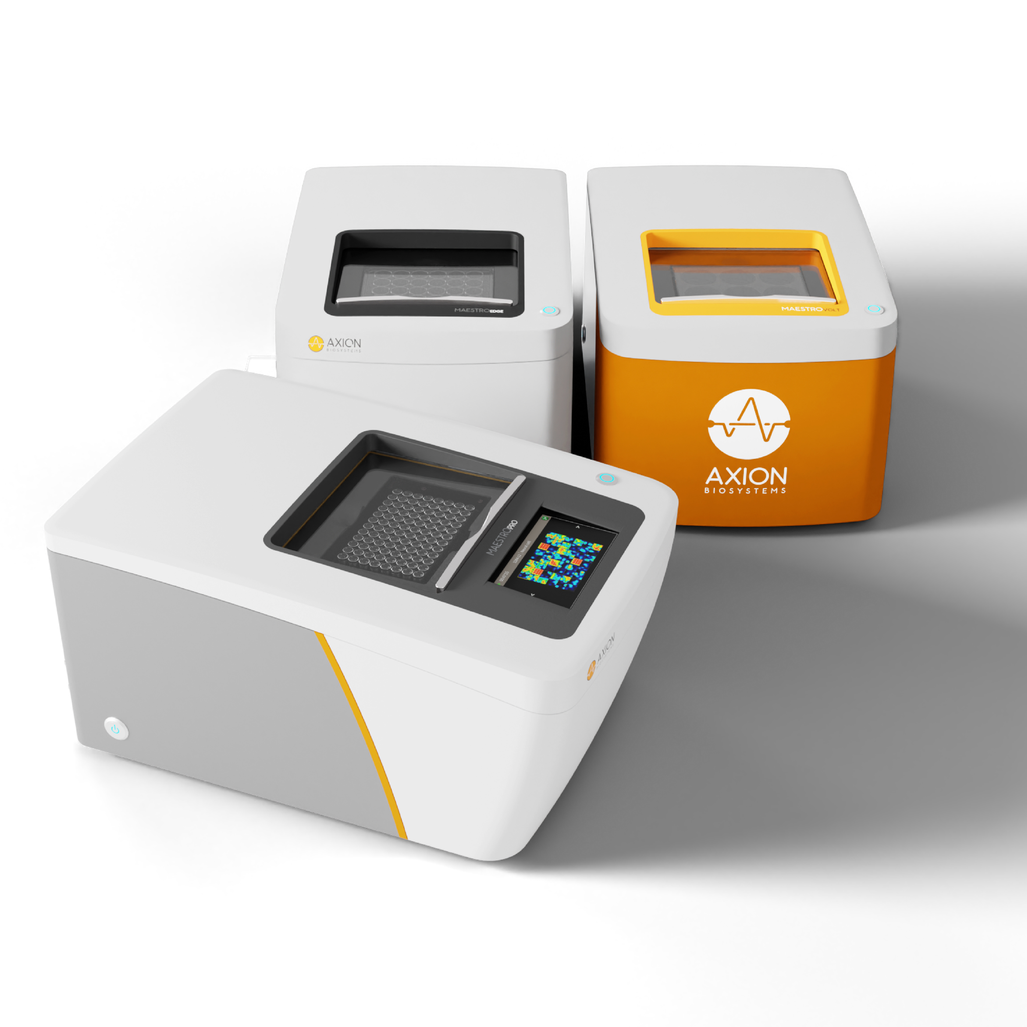
Axion BioSystems offers three microelectrode array systems: Maestro Pro, Maestro Edge, and Maestro Volt, plus a selection of multiwell MEA plates.
Maestro Pro MEA system
The Maestro Pro microelectrode array system is our most advanced offering, designed for the workload of larger labs. Record from our 6-, 12-, 24-, 48-, and 96-well MEA plates.
Maestro Edge MEA system
The Maestro Edge microelectrode array system is a versatile multimodal MEA and impedance system. Record from our 6-, 24-, and 96-well MEA plates.
Maestro Volt MEA system
The Maestro Volt microelectrode array system is our most budget-friendly offering, designed for labs with lower throughput needs. Record from our 6-well MEA plates.
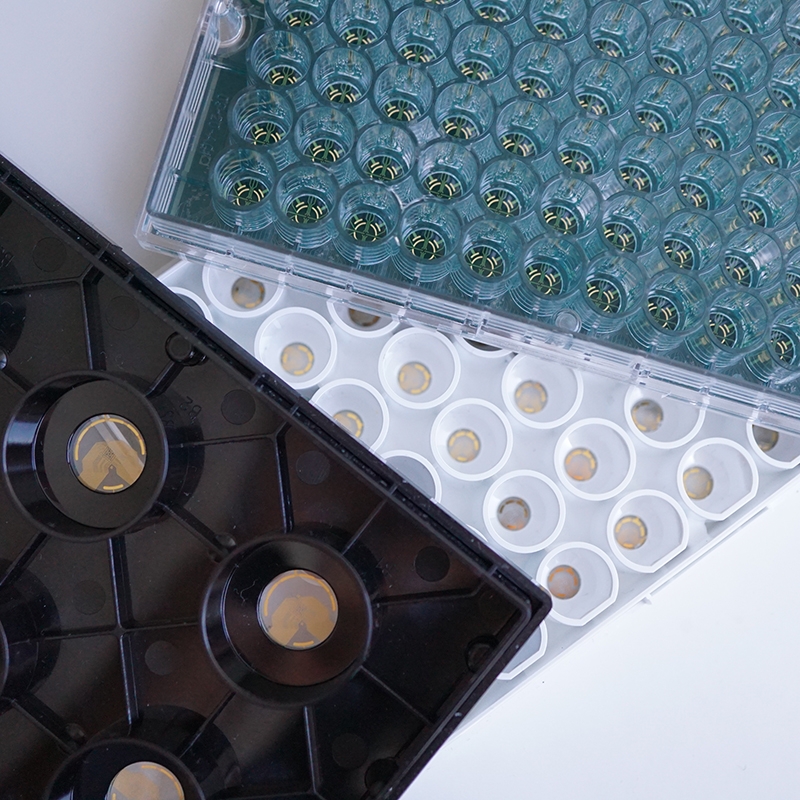
Microelectrode Array Plates
- >> CytoView MEA – The premium Maestro multiwell MEA plate with a transparent well bottom for cell visualization and assay multiplexing. The 24-, and 6-well plate formats are compatible with the Maestro Edge and Maestro Pro.
- >> BioCircuit MEA – Maestro MEA plates with an opaque well bottom delivering high-quality results at the lowest cost per well. The 24-well plate format is compatible with the Maestro Edge and Maestro Pro.
- >> Lumos MEA – Maestro MEA plates designed for use with the Lumos system, featuring a transparent well bottom and light-focusing lid for optical stimulation. The 24-well plate format is compatible with the Maestro Edge and Maestro Pro.
- >> SpheroGuide MEA Plate – Maestro MEA plates with an integrated funnel for accurate placement on the electrodes and a transparent well bottom for visualization. The 48-well plate format is compatible with the Maestro Pro.
Frequently asked questions about microelectrode array?
What is the difference between in vitro microelectrode array and patch clamp?
MEAs record the field potential electrical activity from the extracellular space from a population of electrically active cells such as neurons. Patch clamp electrophysiology records the action potentials from the intracellular space of the neuron.
What are the benefits to using microelectrode array in my studies?
MEAs are high throughput in vitro systems that record from multiple electrodes simultaneously. This can be performed non-invasively, label-free and in real time from cells or organoids. The cells are plated just as they would be in any other multiwell plate.