The development of disorders such as autism and schizophrenia are thought to have their origins in the developing brain. But studying the development of the human brain is challenging due to the absence of good model. In this webinar, Prof. Alysson Muotri (UC San Diego) demonstrates that the spontaneous development of neural networks in neural organoids, commonly referred to as “mini-brains,” resembles those found during early human brain formation. The research findings covered in this webinar were published recently in the journal, Cell Stem Cell, and were featured in numerous media outlets including the New York Times and NPR.
Transcript of webinar on Recording from Neural Organoids on Axion's multi-well MEA system
Thank you for joining for today's Coffee Break Webinar. Today's topic is: A Model of Human Brain Development Measuring Oscillatory Waves in Cortical Organoids.
The development of disorders such as autism and schizophrenia are thought to have their origins in the developing brain. But studying the development of the human brain is challenging. Scientists have up until now relied on animal models, like mice and monkeys, to provide insights into this process. Animal models, however, provide an imperfect representation of the human brain.
The advent of induced pluripotent stem cell technology has provided a readily available source of human brain cells. These iPSC neurons can be grown in a dish and have the benefit of expressing human proteins, such as ion channels and neuroreceptors, with the potential to reflect genetic diversity. However, it’s yet to be demonstrated that these lab grown human neurons recreate the complex neural networks found in the human brain.
In this webinar, Prof. Alysson Muotri, Director of the Stem Cell Program at UC San Diego, demonstrates that the spontaneous development of neural networks in neural organoids, commonly referred to as “mini-brains,” resembles those found in early human brain formation. The research findings covered in this webinar were published last week in the journal, Cell Stem Cell, and has received wide spread interest, being featured in numerous media outlets including the New York Times and NPR.
Thank you for the introduction, Melissa. Hello this is Alysson Muotri, and I am a professor at University of California in San Diego. I am faculty in the Pediatrics department and also in Cellular Molecular Medicine. I’m also the Director of the Stem Cell program here at UC San Diego, and I’ll be talking about applications of brain model technology.
I’ll be focusing on our latest work using brain organoids as a model for neurological conditions. The main motivation for this research is really to create a model to interrogate the electrophysiology of human developing brains. The human brain is really a blank box especially in these very early stages of neurodevelopment. So the access to the human developing brain in uterus is virtually impossible – it would not be ethical to really study these early stages in a healthy human brain. So scientists have heavily relied on animal models or post-mortem tissues to really try to understand how the brain is formed.
So this is a critical step in brain development because that is exactly when the brain is wiring and alterations in these early stages might actually compromise brain function for life. Conditions like autism or psychiatric disorders such as schizophrenia all seem to have origins at these early stages. So understanding how the brain is wired at these very early stages is quite critical to understand how the brain works.
Brain organoids generated from induced pluripotent stem cells, or iPSCs, have emerged as a scaled down and three-dimensional model of the human brain, mimicking various developmental features at the cellular and molecular levels. And despite recent advances in the understanding of the cellular diversity, there is no evidence that these organoids can actually form complex and functional neural networks that might resemble early human brain formation.
So our research has been focusing on trying to see if these brain organoids can actually mimic these very early stages of neural network formations. In brain organoids, as is in any other models, they actually have their intrinsic limitations.
Most of those neurons are immature neurons. The organoids are not vascularized. We don’t have all cell types represented. We don’t even know if we are growing them in the ideal culture conditions. So there are several questions regarding these limitations which are fair questions. For example, are they a good model to show translatability. Is this translational? So we are pushing this field to actually have a model where we can study these brain organoids not only at the molecular and cellular level but also at the networks dynamics.
We ask if this technology can be disruptive on helping us to really understand how the brain networks mature over time. To further evaluate the cortical organoids functionality at a mesoscopic level, we perform weekly extracellular recordings of spontaneous electrical activity using multielectrode arrays or MEA over the course of 10 months.
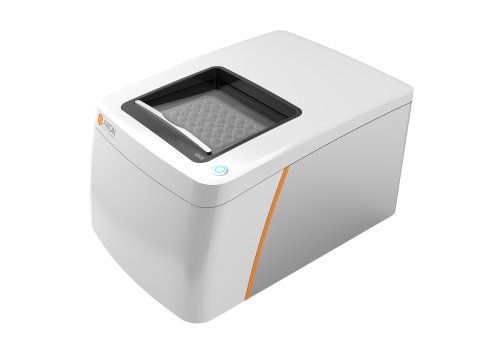
Cortical organoids were plated in wells of these MEA plates. Each well containing 64 low impedance platinum microelectrodes using a total of 512 channels. So we virtually generate activity maps as well as raster plots every week so we could analyze how the electrical activity would evolve over time. Over the course of 10 months, the cortical organoids exhibit consistent increases in electrical activity.
We can measure that by channel-wise firing rate, burst frequency and synchrony. Which indicates a continuously evolving neuro network. Additionally, the variability between the replicates over the 40 weeks of differentiation was significantly lower compared to the iPS-derived neurons in monolayer 2D cultures. Which was a big surprise to us.
During the initial recordings, we noticed that our organoids displayed a very robust pattern of activity. Reaching between long periods of quiescence where the networks were virtually silent with short bursts of spontaneous network synchronized spiking, or network events. These network events were periodic and infrequent early on in development when they’re about two months. They were happening every 20 seconds in decay after the initial onset.
When they reach about 4 months, we can see a second peak emerging after the initial network event leading to the presence of what we call a nested faster oscillatory pattern between two and three Hertz. They went up to 6 months in culture. So this is a robust, fast, nested oscillation, cannot be seen in a 3D neuro sphere for example. So it’s not a matter of number of neurons in a 3D configuration.
So it only really happens when you grow these organoids from single cells letting these neurons to form these network activities over time. So it seems to us that as long as you let them do what they are supposed to do, the genetic information codes for these kind of network to happen. So we quantify this complexity using several measures both spatial and temporal correlations between network events. For example, in inter-event interval consistently increase over 10 months of differentiation, from extremely regular latencies at 2 months to irregular at 10 months. So this indicates that increasing variability between consecutive networks events increase over time as we would expect for a more complex network.
The spatial and temporal irregularity at the short term time scale within events also increase with development. Suggesting a breakdown of the deterministic population dynamics from the onset of the network events. This level of activity and this oscillatory behavior was never seen in tissue cultures very unprecedented especially for human iPS-derived neurons. So we are curious to see these complex oscillatory network activities in these organoids organoids can actually represent the spontaneous developmental trajectory observed in early human neurodevelopment.
So while the network activity for cortical organoids does not exhibit the full temporal complexity seen in adults, the pattern of altering periods of quiescent silent time in network synchronized events resemble the electrophysiological signature present in pre-term human infant EEGs or electroencephalograms. This is what we call the trace discontinue. So quiescent periods punctuated by high amplitude oscillatory behavior lasting just a few seconds. These intervals of complete quiescence disappear as infants become of term in the EEGs dominated by continuous low amplitude desynchronized activity in adult brains. So we thought that by using pre-term EEGs would be a nice way to start to correlate our own data with human neurodevelopment.
I think it’s important to mention that the biophysics of the scalp EEGs is dramatically different from extracellular field potential in the cortical organoids. Mainly because of factors such as spatial filtering by the scalp, or the orientation of neuronal populations in relation to the recording electrode. So therefore we have to select the features that are definitely comparable between the two systems. That’s how we decided to select only a few of them to actually compare the cortical organoids in the preterm neonate EEGs.
By comparing specific timing features between cortical organoids and the preterm infants, we found a range of correlations in the developmental trajectory of features with age, as well as with similarities in development between the two datasets. So our regression model predicted organoid development time very poorly before 25 weeks. We have high variability followed by a true age with higher fidelity after 25 weeks. And the reason is that we could not train the machine before 25 weeks. There is really not much data on preterm EEGs from earlier than 25 weeks. So a subset of preterm EEG held out during training and was used for further validation of the model.
In addition, we use other controls such as mouse primary culture, iPS monolayer cultures, and even human fetal brain cultures. So it’s only the brain organoids whose showed these nice correlation after 25 weeks. So note that a significant positive correlation was only observed in the organoids and held out in EEG datasets. So while the developmental trajectory of the cortical organoids is definitely not identical, and are more variable than what we see in the human fetal brain, the two populations share similarities in how their networks electrophysiological properties, change over time. Suggesting a genetically programmed developmental timeline that can be detected by a simple machine learning algorithm.
So future research using this model. My lab has been focusing on recent clinical trials for autism using CBD, cannabidiol, where we are generating a clinical trial in vitro in parallel. Every subject in this clinical trial will have their brain organoids made and measured using the same technology. What are hoping is to see how CBD can change the electrical properties of these oscillatory waves in these brain organoids, hoping to predict or inform the clinical data beforehand. If that’s true, it would show a validity of this in vitro model to actually correlate at least partially some of the features observed in the brain organoids with the actual subjects. We are really exciting in waiting for this data to come. We just started, and this is something to watch in the next year or so.
The data really shows spontaneous development of neural networks in brain organoids. In using these simple machine learning algorithms, we show that they do have something that seems like a genetically encoded biological program because the trajectory between the organoid and the human preterm baby seems very similar. We detected these delta-high gamma phase-amplitude coupling during these network synchronous events. Which is again, a hallmark of inter-regional cortical communication. This is suggesting that probably what we are having in these organoids to have such a high level of activity nested oscillatory behavior is some kind of micro-circuitry is being formed as we let them mature.
The organoid electrophysiological signature mimics the preterm neonatal brain especially after 25 weeks post-conception. We can not train the machine before that, just because of the lack of data. The potential use of these brain organoids to model neurodevelopmental and disease states are ongoing as I mentioned. These clinical trials are something that really show validity on translatability as one of the key features that we think that these brain organoids might be useful in the future. I hope this was useful. There are tons of data in the manuscript. I invite you to read, comment, send me messages if you have any questions.
This work was led by three researchers in my lab. This is Cleber Trujillo, whose really a leader in this field now, and has been pushing a lot to get this model going. Priscilla Negraes is another post doc in the lab, and a grad student in the Voytek lab, Richard Gao. These three actually did the vast majority of the ground breaking work that you see in this manuscript. My funding agencies, the NIMH and CIRM, they heavily contributed to create this new model here. And I need to further disclosure here that I am a cofounder of TISMO. This is a company that is using this technology of brain cortical organoids for drug screening for autism and other neurological conditions. And finally, all the families and subjects that contributed to our research.
If you want to learn about how to record from Mini-brains on Maestro MEA visit axionbio.com/mini-brain for more details.
And that is the conclusion for today’s Coffee break webinar. If you have any questions you would like to ask regarding the research presented or if you are interested in presenting your own research with microelectrode array technology, please forward them to coffeebreak@axionbio.com. For questions submitted for Dr. Muotri, he will be in touch with you shortly.
Thank you for joining in on today’s coffee break webinar and we look forward to seeing you again.


