Lumos OptiClear Plate
The Lumos OptiClear™ plate has a high clarity bottom culture surface for microscopy and high content imaging. Available in 24-, 48- and 96- well options, the Lumos OptiClear plates have specially formulated reflective white walls and lensed lid facilitate optimal light delivery within each well, with minimum crosstalk between wells.
Key Features
High throughput optical control.
- High throughput, multiwell format: 24-, 48-, and 96 wells of simultaneous light stimulation.
- Optimal light delivery and dispersion: Customized plate material and lid optics enhance light delivery and allow for even light dispersion. Use with Axion's Lumos system to perform optogenetic experiments.
LUMOS OPTICLEARThe Lumos OptiClear plates for imaging assays with white walls and custom optical lid for optimal light delivery in each well | |||||||||||
|---|---|---|---|---|---|---|---|---|---|---|---|
| Plate | Cat No. | Wells | Electrode/ | Electrode | Bottom | Walls | Maestro Volt | Maestro | Maestro | Maestro | Maestro |
| Lumos OptiClear 24 | OPT-24 | 24 | 0 |  | Transparent | White | |||||
| Lumos OptiClear 48 | OPT-48 | 48 | 0 |  | Transparent | White | |||||
| Lumos OptiClear 96 | OPT-96 | 96 | 0 |  | Transparent | White | |||||
*Schematic of well illustrating that there are no recording electrodes in Lumos OptiClear plates. Consequently the Lumos OptiClear plates are not compatible with any of the Maestro systems. Lumos OptiClear plates are intended for use with the standalone Lumos optical stimulation system and in downstream imaging or plate-reader assays.
Overview
High quality optical performance
The Lumos OptiClear plates are composed of custom-formulated plate walls that provide high reflectance to maximize light delivery to your culture and minimize well-to-well crosstalk. The custom-molded lid mates to the Lumos array and contains integrated optical lenses. Finally, the transparent bottom allows for cell visualization and assay multiplexing with fluorescence, luminescence, and other reporter-based assays.
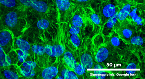
QBM rat cortical neurons fixed on 14 DIV and stained for nuclei (blue, DAPI) and F-actin (green, phalloidin). Image courtesy of the Santangelo Lab, Georgia Tech.