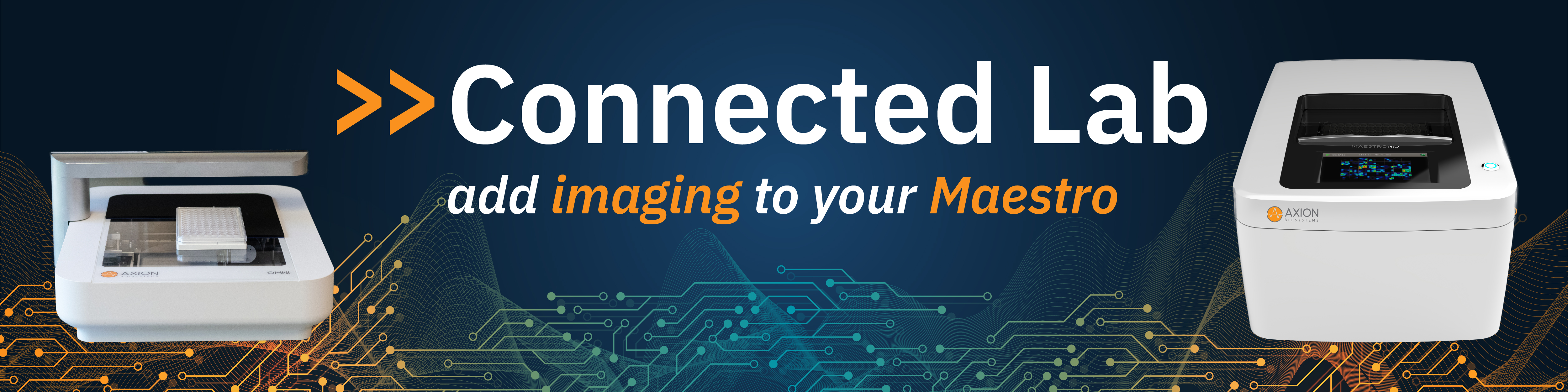
Seeing is believing
Discover the power of the Connected Lab and get a more comprehensive view of your 2D and 3D in vitro cell models with side-by-side imaging and MEA data.
- >> Correlate function & morphology
- >> Improve data interpretation
- >> Monitor culture quality over time
- >> Ensure even, consistent coverage
Visualize your cells in AxIS software with no complicated steps. Axion Portal (included with Omni) automatically links plates via barcode so whole-well imaging data will appear in AxIS Navigator.

Optimizing MEA assays with imaging
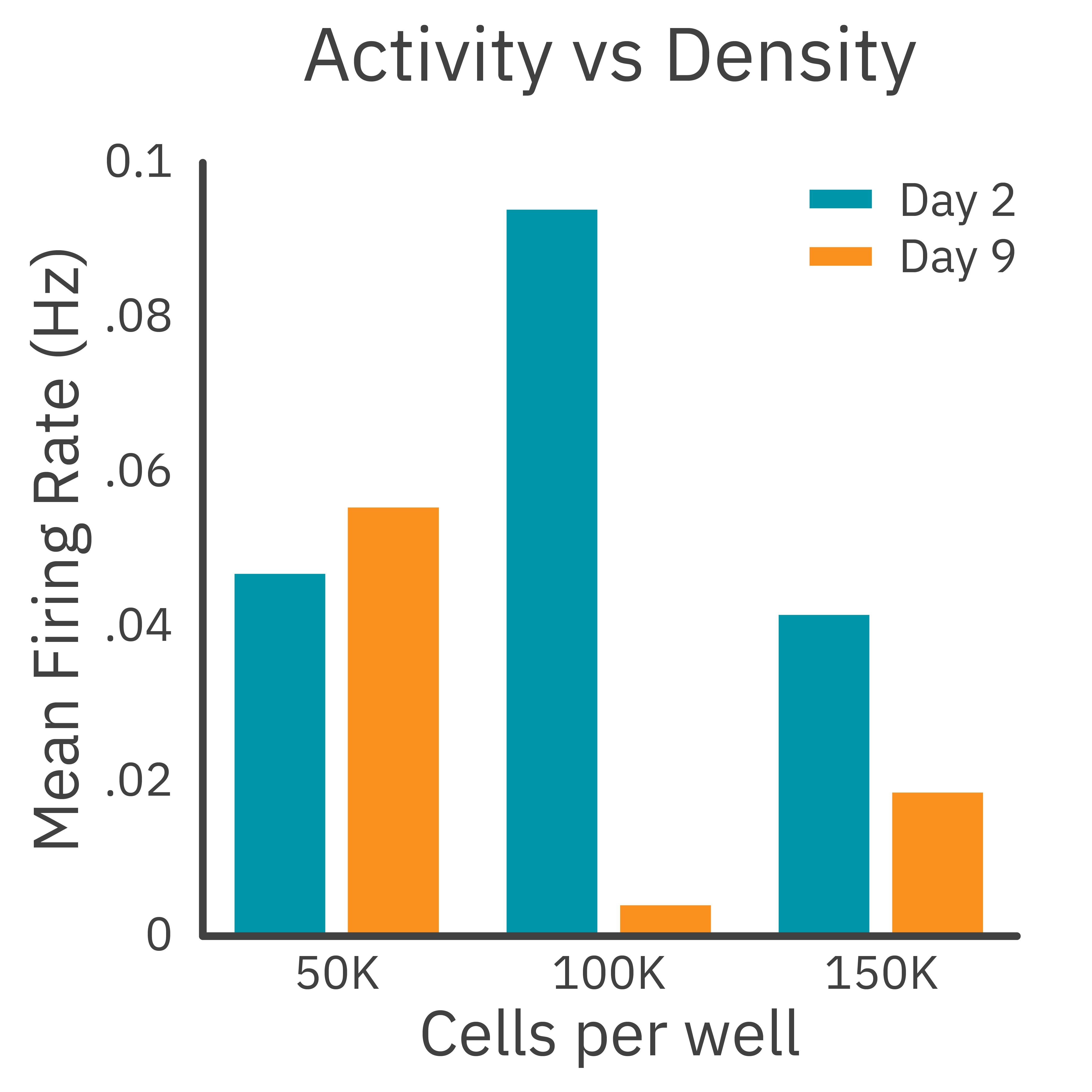
Finding the right density and timing is critical for your MEA experiments. Visual confirmation of cell attachment and distribution ensures reliable recordings and reduces experimental variability.
In this case study, LUMC researchers aimed to optimize the density of new cell lines on the Maestro MEA. While activity showed a drop-off at the highest density and a decrease over time in the medium density, live-cell imaging revealed that higher densities led to increased cell clumping and poor attachment.
With the Connected Lab, the team was able to simultaneously optimize cultures for even coverage and activity.

Data courtesy of Harald Mikkers, PhD,
Dept. Cell Chemical Biology,
Leiden University Medical Center (LUMC)
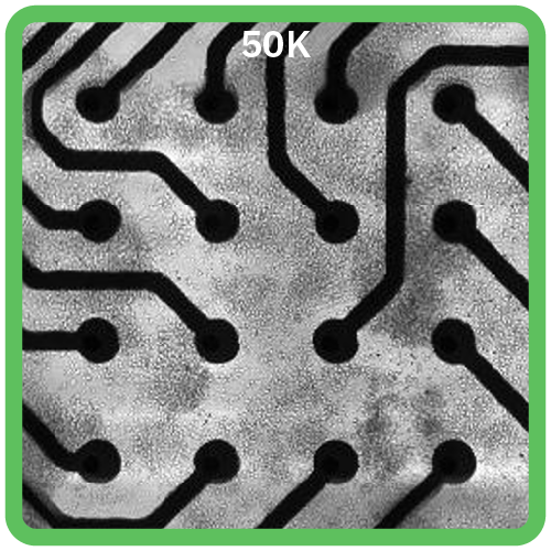
Optimized
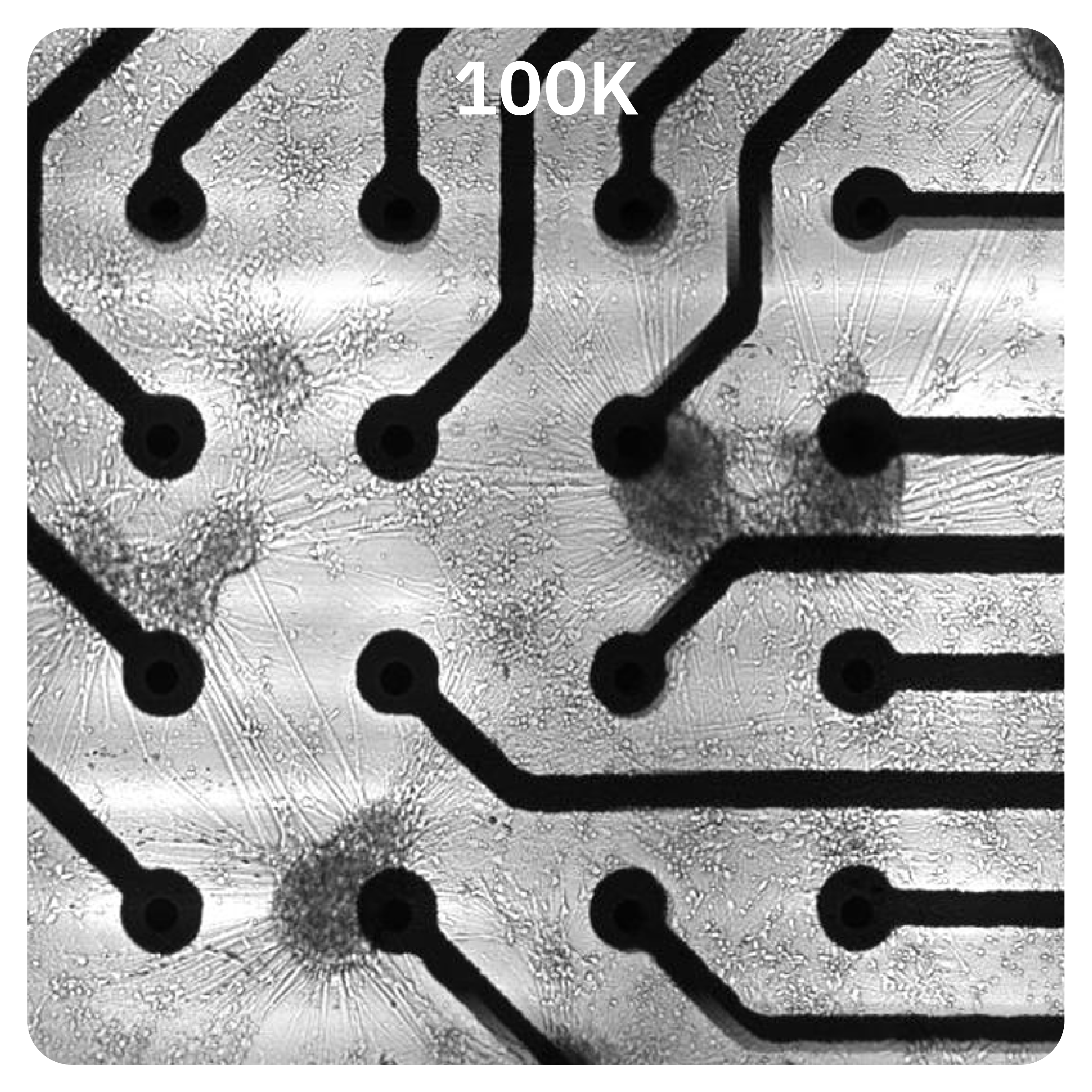
Sub-optimal
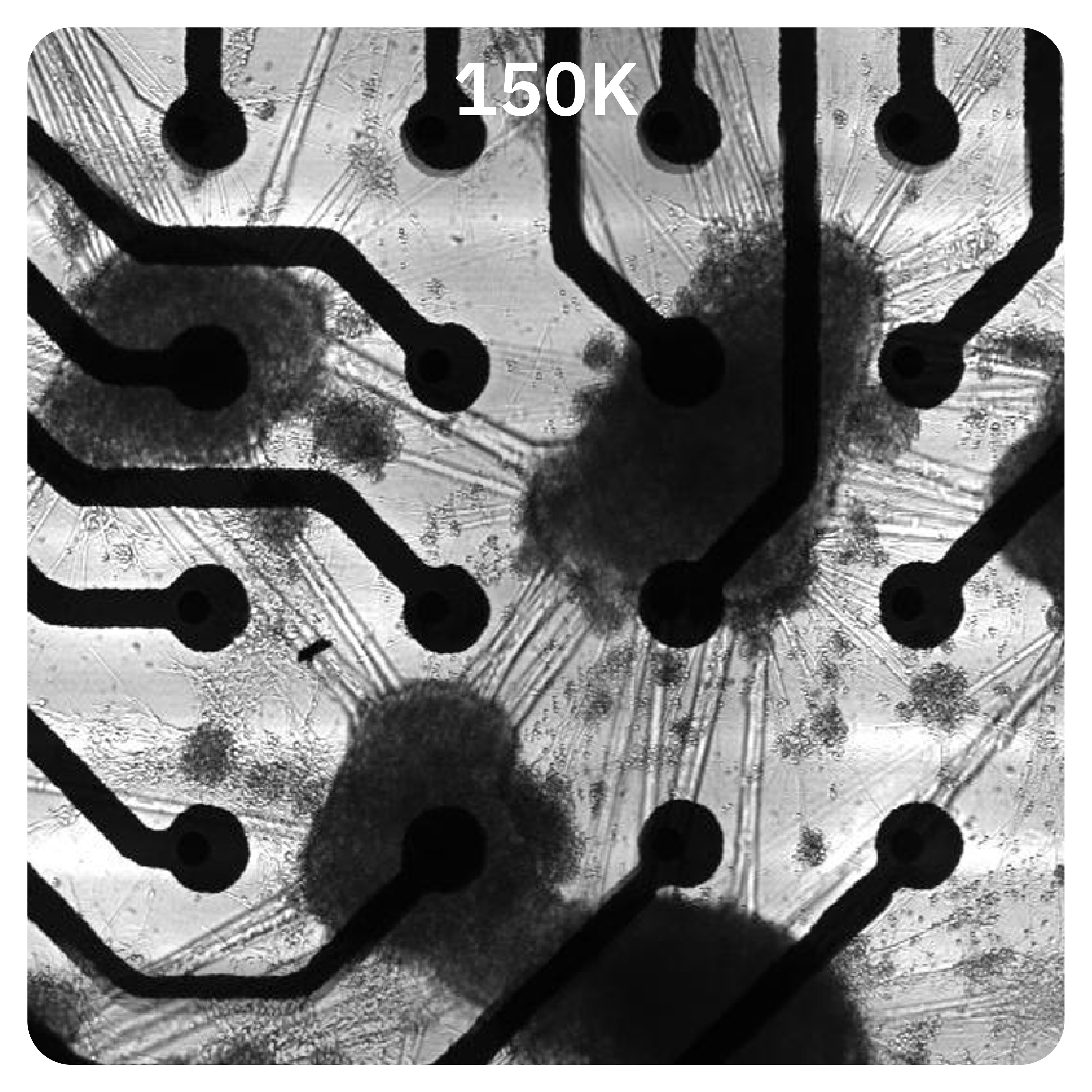
Sub-optimal
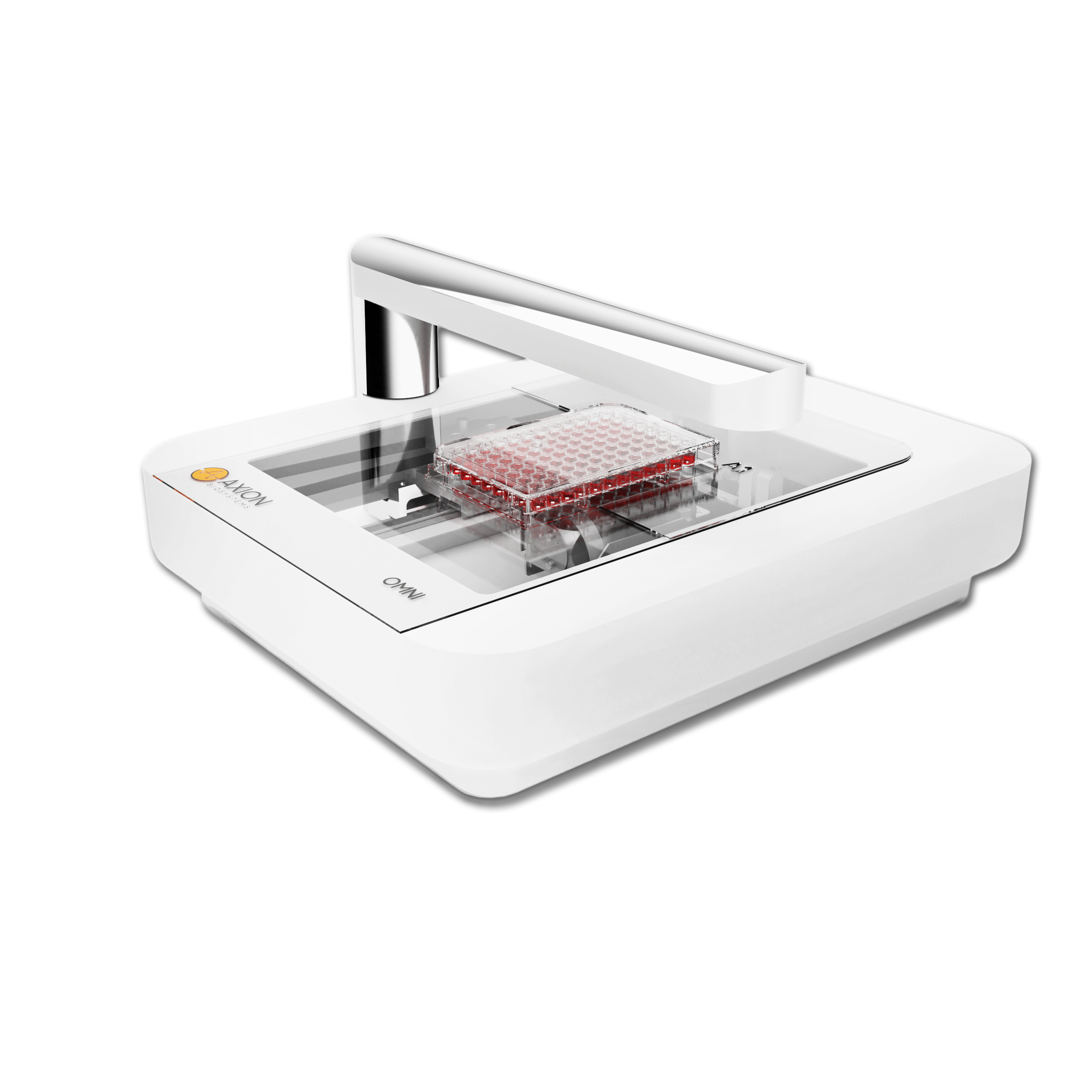
Streamline your MEA with the essential live-cell imager
The Omni imager adds dynamic visual results to any experiment. Featuring AI-supported analysis, brightfield and fluorescent capabilities, and the versatility to scan all types of culture vessels from your incubator, it’s the perfect match for your Maestro MEA experiments.
Speak to our specialist
Learn how to get the “complete picture” with the Connected Lab.