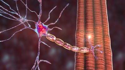
Amyotrophic lateral sclerosis (ALS) is a neurodegenerative motor neuron disease that can be modeled in vitro to study how genetic mutations, protein misfolding, neuroinflammation, and other disease-associated mechanisms alter cellular function. For researchers developing ALS disease models, functional readouts such as neuronal firing, bursting, synchrony, and network activity can provide important insight into how disease biology affects motor neuron behavior over time.
Axion’s Maestro Microelectrode Array (MEA) system, enable noninvasive measurement of electrical activity from neurons in vitro, helping life science researchers characterize ALS-associated functional phenotypes and evaluate responses to experimental treatments in cell-based models.
In Vitro ALS Model Characterization
Amyotrophic lateral sclerosis (ALS) is a neurodegenerative disease often modeled in vitro using iPSC-derived motor neurons, neuromuscular junction co-cultures, and related cellular systems. For researchers, functional phenotypes such as hyperexcitability, firing rate changes, burst patterns, and network activity provide measurable readouts for disease biology and therapeutic screening.
Axion’s in vitro Maestro MEA assay platform allow researchers to observe the decline in neural function associated with the development of ALS.
Study Neuropathology of ALS Using in vitro ALS Models
-
Identify phenotypic differences of ALS motor neurons>
-
Test the efficacy of potential ALS treatments in vitro>
-
Model neuromuscular junctions and stimulate with light>
-
Publication Highlights: Amyotrophic lateral sclerosis (ALS)>
Purpose: To evaluate in vitro phenotypes of iPSC-derived ALS motor neuron models. It has previously been reported that ALS neurons demonstrated neural hyperexcitability in vivo.
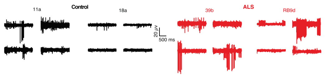
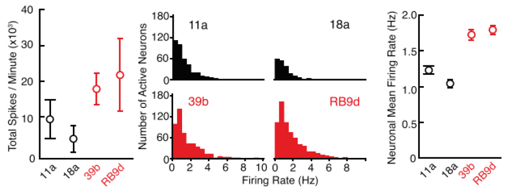
Activity of iPSC-derived ALS neurons harboring SOD1A4V/+ mutation and control cells was measured using patch clamp and the Maestro MEA platform.
Result: ALS neurons demonstrated an increase in firing in both patch clamp and MEA, suggesting that ALS neurons are more hyperexcitable compared to the control cells as observed in in vivo models. Because MEA can measure from an entire culture in higher throughput, researchers used it to confirm the phenotype across mutations. [Wainger et al. 2014].
Purpose: To validate drug screening results to reverse ALS hyperexcitability phenotype. After MEA confirmed hyperexcitability across all tested ALS-derived neurons, the researchers began to test compounds to reverse this phenotype.
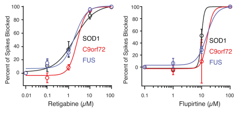
Potassium channel openers retigabine and flupirtine were tested on iPSC-derived, ALS neurons, and the firing rate was measured on the Maestro MEA platform.
Result: Both drugs were found to reduce ALS-related hyperexcitability. As a result of these studies, Retigabine entered into clinical trial as a potential treatment for ALS. [Wainger et al. 2014].
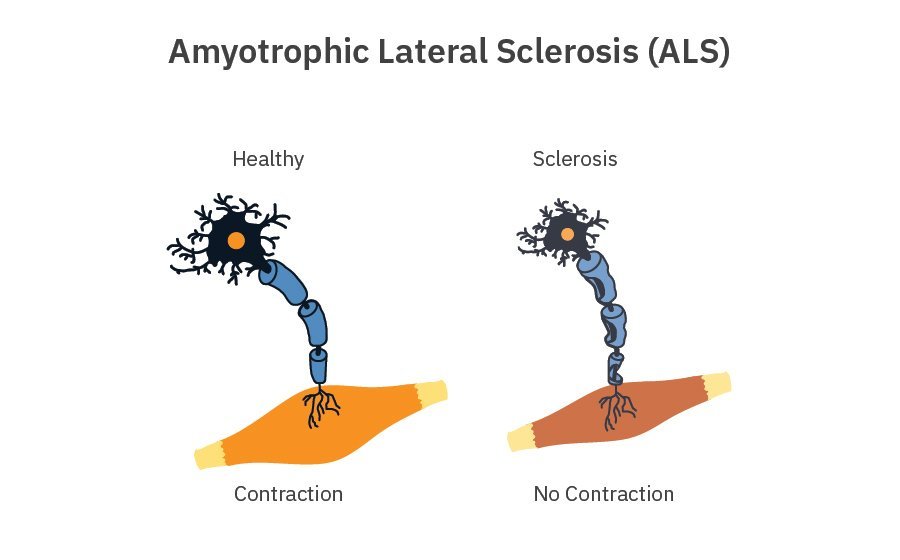
Purpose: to create an in vitro model of the neuromuscular junction (NMJ). The failure of the NMJ is a key component of degenerative neuromuscular diseases, including ALS. A co-culture model may offer patient-specific significant insights into pathogenic mechanisms that underlie NMJ dysfunction in disease.
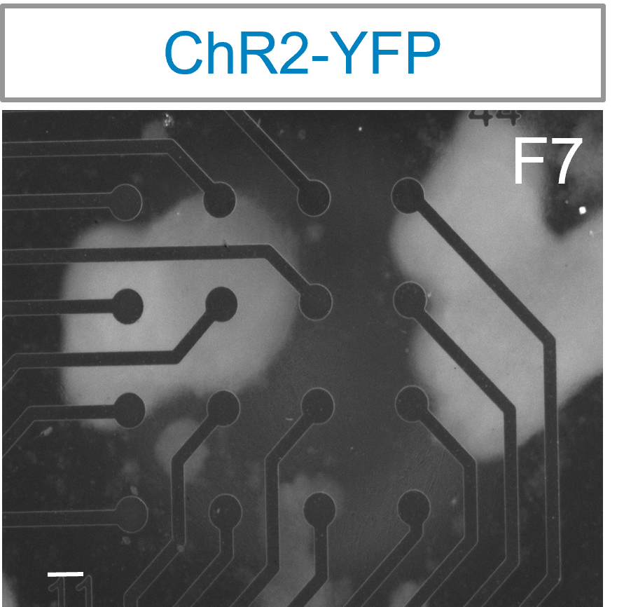
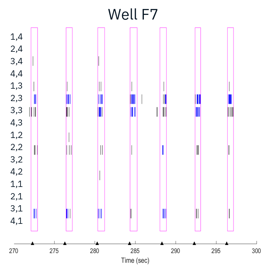
Channel rhodopsin (ChR2) expressing motor neurons were cocultured with skeletal myotubes. Motor neurons were able to elicit muscle contractions when stimulated with blue light using the Lumos.

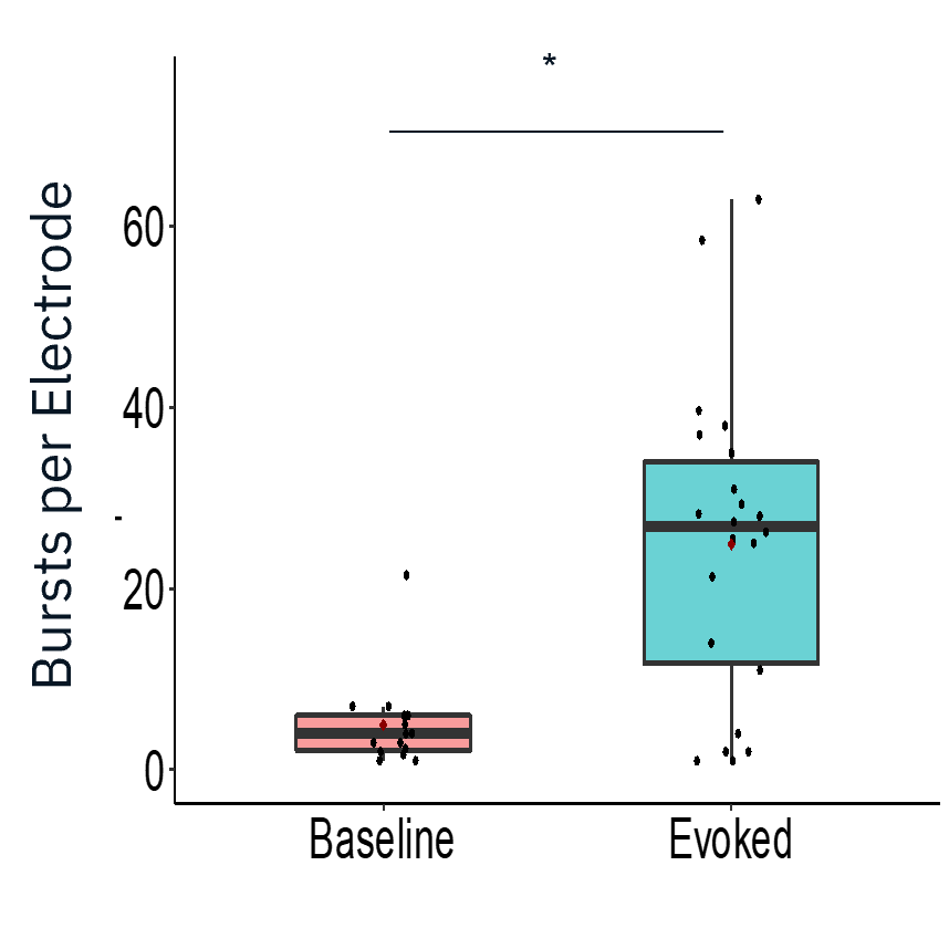
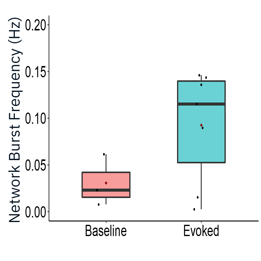
Characterization of evoked firing activity using the Maestro Pro showed robust evoked activity.
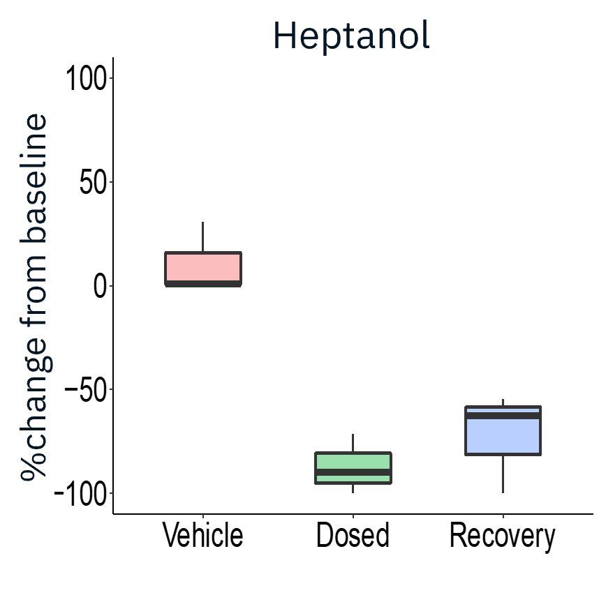
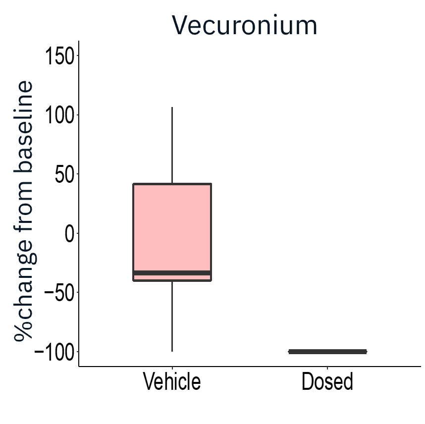
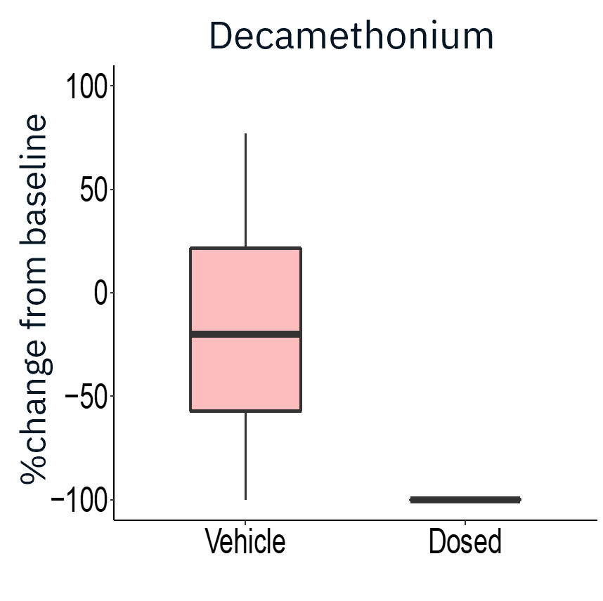
The application of gap junction blocker heptanol or neuromuscular antagonists decamethonium bromide and vecuronium drastically reduced evoked activity, demonstrating that activity measured was primarily from skeletal myotubes.
Result: The Maestro MEA platform could selectively stimulate motor neurons which activated skeletal myotubes, demonstrating a functional neuromuscular junction that responded appropriately to compounds.
Publication Highlights: Amyotrophic lateral sclerosis (ALS)
Learn how Axion's live-cell platforms were used in ALS research with these selected publications.

Frequently Asked Questions: In vitro ALS Disease Model MEA Assays
Microelectrode array (MEA) technology enables noninvasive measurement of electrical activity from cultured neurons over time. In ALS models, researchers can use MEA to assess functional phenotypes such as neuronal firing, bursting, synchrony, network activity, and changes in activity following experimental treatment.
- The Maestro MEA platform offers a controlled environment for studying detailed neural network activity in vitro.
- High-throughput multi-well plates make it ideal for screening patient-specific lines and therapeutics.
- Noninvasive monitoring allows for the study of long-term effects and disease progression.
- It is easy to use, requiring only basic cell culturing techniques to measure neural electrophysiology.