
Lumos MEA 플레이트
Axion BioSystems 의 Lumos multiwell MEA 플레이트로 고도로 최적화 된 광학 성능과 함께 높은 수준의 MEA 데이터를 확보할 수 있습니다.
24, 48, 96-well 중 선택할 수 있으며 Axion 에 맞춰 제작된 Lumos 리드와 특별 제작된 플레이트는 well 간의 간섭현상은 최소화하면서 광자극 전달은 최대화 하도록 최적화되어 있습니다. 또한 투명한 well 바닥은 세포의 시각적 관찰과 형광, 발광 또는 그외의 다른 리포터 기반 연구들의 동시 수행을 지원합니다.
Key Features
고처리량의 광학 조절
-
고처리량의 Multiwell 타입 - Lumos MEA 플레이트는 높은 수준의 데이터 확보가 가능합니다.
-
광자극 전달 및 산란에 최적화 - 맞춤형 플레이트와 리드는 광자극 전달을 향상시키고 균일한 빛 산란에 적합합니다.
-
다양한 실험 동시 수행 - Axion 의 Lumos 시스템의 광유전학 실험은 MEA 데이터를 한층 보완하는 데 활용할 수 있습니다.
Lumos MEAThe Lumos MEA plates combine robustness and assay flexibility of a CytoView MEA plate with white walls and a custom optical lid for optimal light delivery in each well
|
||||||||||
| Plate | Cat No. | Wells |
well |
layout* |
|
|
Edge |
Pro |
Z/ZHT |
Original |
|---|---|---|---|---|---|---|---|---|---|---|
| Lumos MEA 24 | M384-tMEA-24OPT |
|
|
 |
|
|
|
|
|
|
| Lumos MEA 48 | M768-tMEA-48OPT |
|
|
 |
|
|
|
|
|
|
| Lumos MEA 96 | M768-tMEA-96OPT |
|
|
 |
|
|
|
|
||
*Schematic of well illustrating recording electrodes (blue), grounds (orange), and where present, a large dedicated stimulation (blue).
Overview
세포의 시각적 관찰과 다양한 실험수행
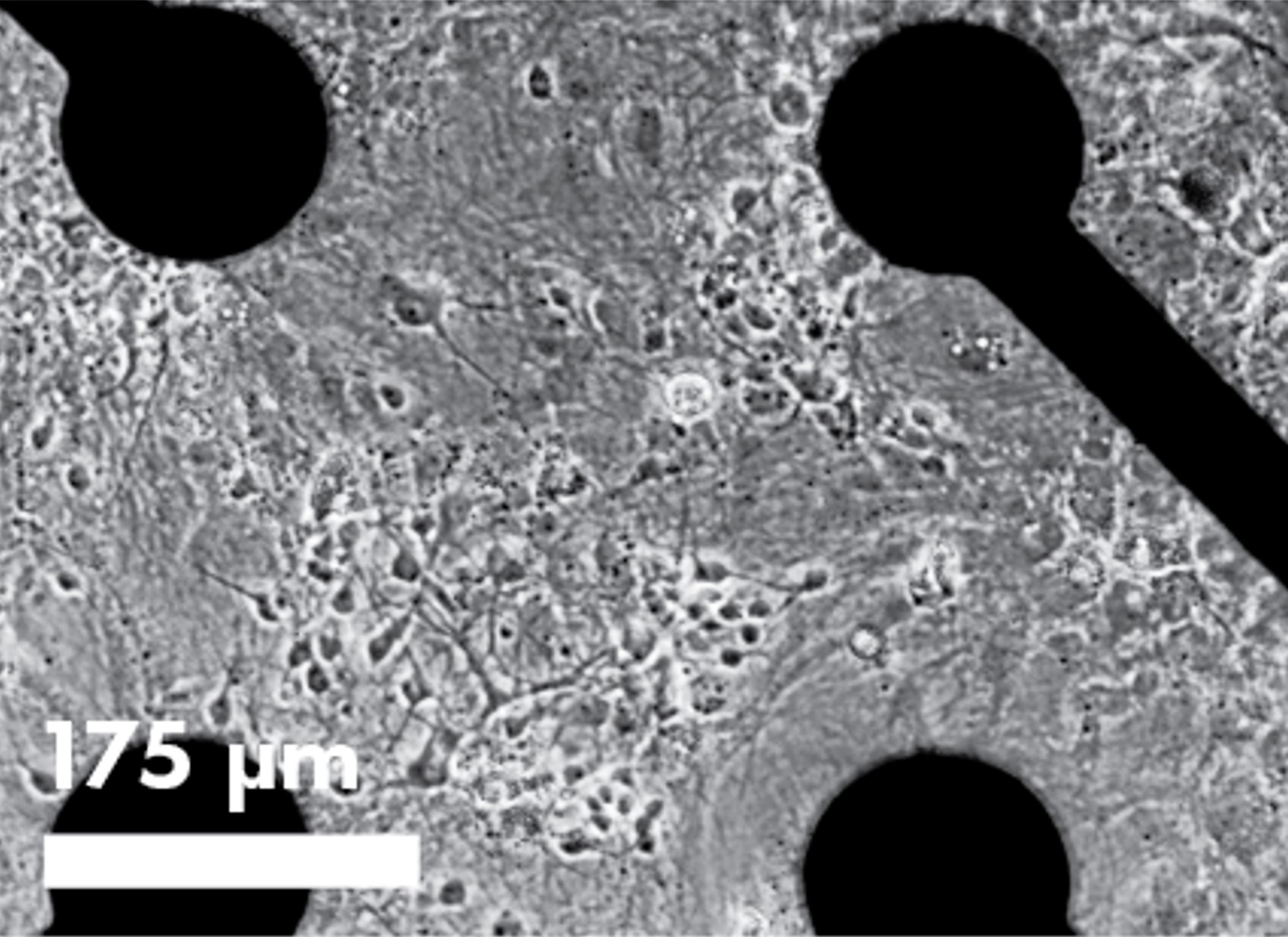
혁신적인 투명한 플레이트 바닥은 세포 시각화 및 여러 실험을 동시 수행할 수 있도록 지원합니다. 명시야 이미징은 세포가 정확한 위치에 자리잡았는지 확인할 수 있도록 도와주고 세포가 건강하게 서로 연결되었을 때 보여지는 MEA 결과에 신뢰를 더해줍니다. 또한 여러 형광 혹은 발광 실험과 같은 End point 실험 결과로 MEA 측정 결과를 보완할 수 있습니다.
광유전학적 접근으로 세포의 파열(bursting) 혹은 조율(pacing)을 제어해보세요
로돕신(ChR2)와 같이 빛에 민감한 흥분성 이온 채널을 발현하도록 유전학적으로 변형된 세포를 이용하여 심장세포 박동 조율 혹은 신경 세포 네트워크 파열을 제어할 수 있습니다.
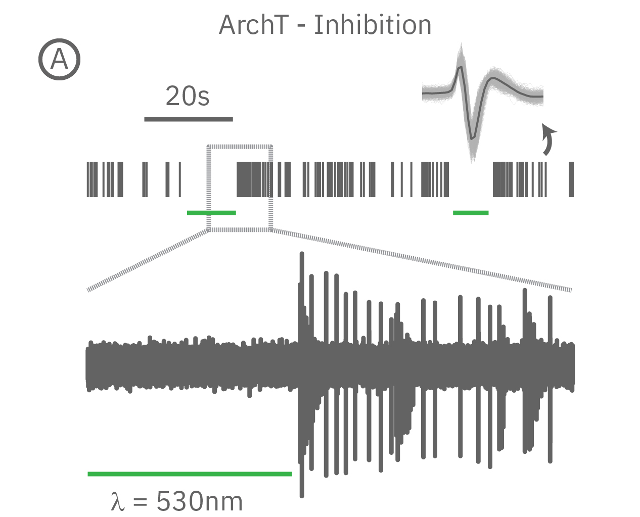
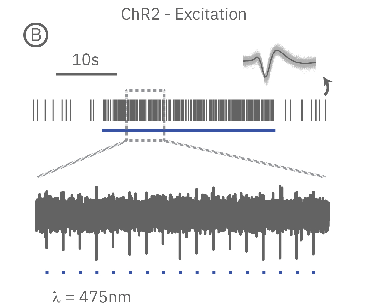
(A) When expressed in neurons, ArchT suppresses neural activity upon incident green light. (B) Whereas, ChR2 can be used to activate neurons in vitro in response to blue light.
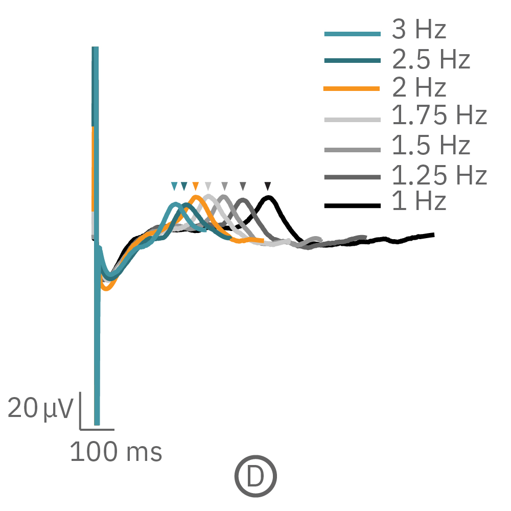
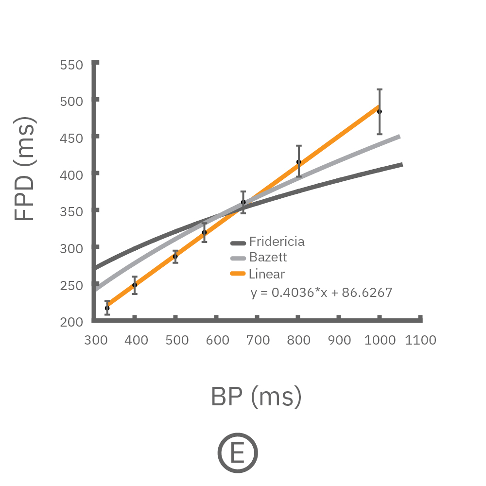
심장세포의 경우, 심장의 재분극은 약물 조절에 민감한 심박동 주파수와 본질적으로 관련이 있습니다. 광유전학적 자극은 심박동 주파수를 제어함으로써 변수를 제거하고 재분극 측정 결과의 신뢰도를 높이는 데 활용할 수 있습니다.
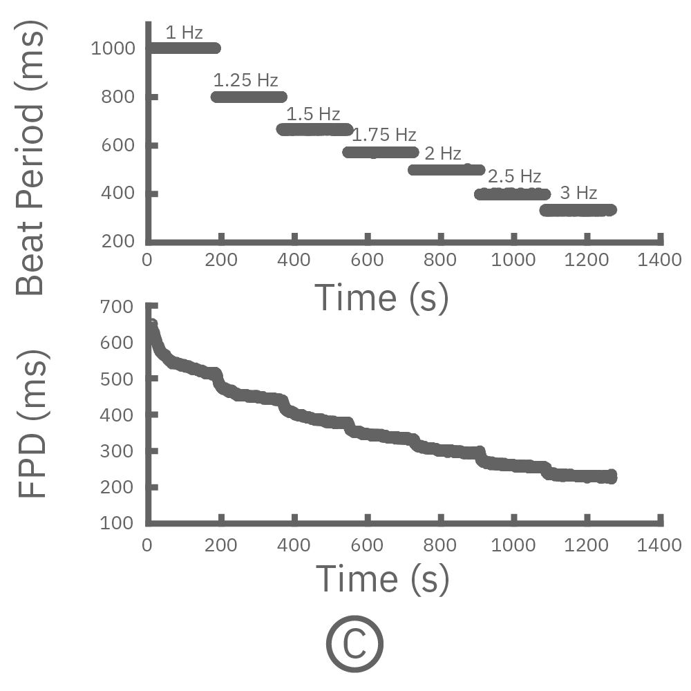
(C, D) The beat rate of Pluricyte® Cardiomyocytes was increased in a step-wise manner (known as a “chirp” assay). The field potential duration (FPD) adapted with each sequential beat rate increase up to 3 Hz. (E) Typical clinical correction formulas, the Fridericia and Bazzett, did not accurately predict the FPD. However, pacing with the Lumos revealed the cell-specific beat rate correction relationship.